![]()
Abdominal CT: appendicitis
Identifying acute appendicitis
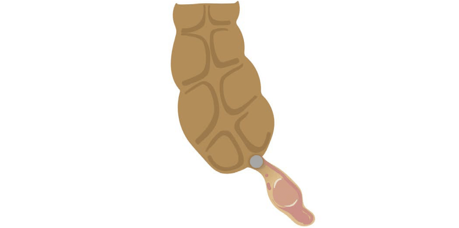
Acute appendicitis occurs when the connection between the appendix and cecum becomes blocked, often by a small stone called an appendicolith or an overgrowth of lymphoid tissue. This causes fluid to build up in the appendix, resulting in inflammation and infection, and can potentially cause a loss of blood flow and rupture of the wall.
Acute appendicitis is a common indication for abdominal computed tomography (CT) in younger patients presenting with fever, right lower quadrant pain and elevated white blood cell count. Acute appendicitis is also an important diagnosis to consider in older patients, although it is less common in this group.
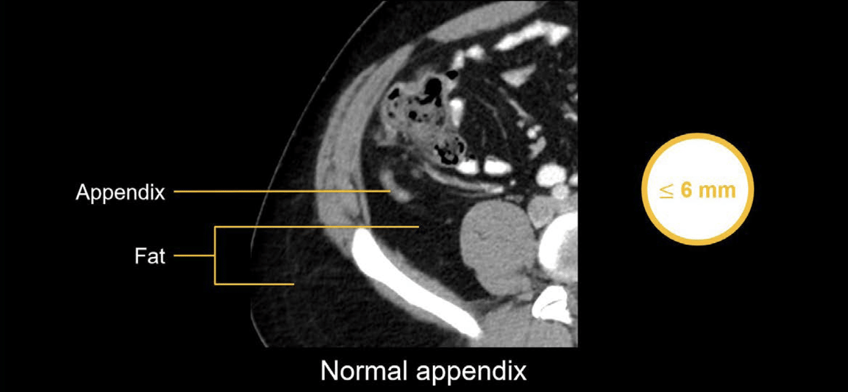
Normal appendix
The normal appendix is usually 6mm or less in diameter and is associated with a clean or dark appearance of the surrounding fat. In the image shown next, notice how the fat surrounding the appendix looks similar to the body wall and other areas in the abdomen.
Acute appendicitis
Three key imaging findings can help you confidently diagnose acute appendicitis:
- A dilated, fluid-filled appendix that is greater than 6 mm in width
- Inflammation around the appendix (i.e., stranding or wavy lines and haziness of the fat surrounding the appendix)
- Bright, calcified deposit(s) in the appendix (i.e., appendicolith) which can obstruct the lumen and be the cause of appendicitis

Clinical Case 1
Try out an evaluation of the appendix using our online PACS viewer. This case involves a 25-year-old with right lower quadrant pain.
- Pull up both the axial and coronal images side-by-side to give you the best view of the anatomy
- The patient received both oral and intravenous (IV) contrast, so notice that you can see bright contrast within the small bowel loops.
- Scroll down to the right lower quadrant and identify key anatomic landmarks: – ascending colon – cecum – ileocecal valve – terminal ileum
- Notice the dilated, tubular, and rather angry-looking structure behind the cecum.
- Scroll through the structure on both the axial and coronal images, tracing it back to the cecal base and confirming that it is the appendix.
- Notice the thickened wall of the appendix and typical stranding and haziness of the surrounding fat, indicating inflammation. This is a typical look for acute, uncomplicated appendicitis which can be confidently diagnosed in this case.
Perforated appendicitis
When the appendix is perforated (i.e., ruptured), many people expect to see a straightforward, well-defined perforation with air and fluid outside of the appendix. While that may be the case with some patients, there is a spectrum of different presentations we can see from early perforation to later perforation. The stage will greatly influence what you see.
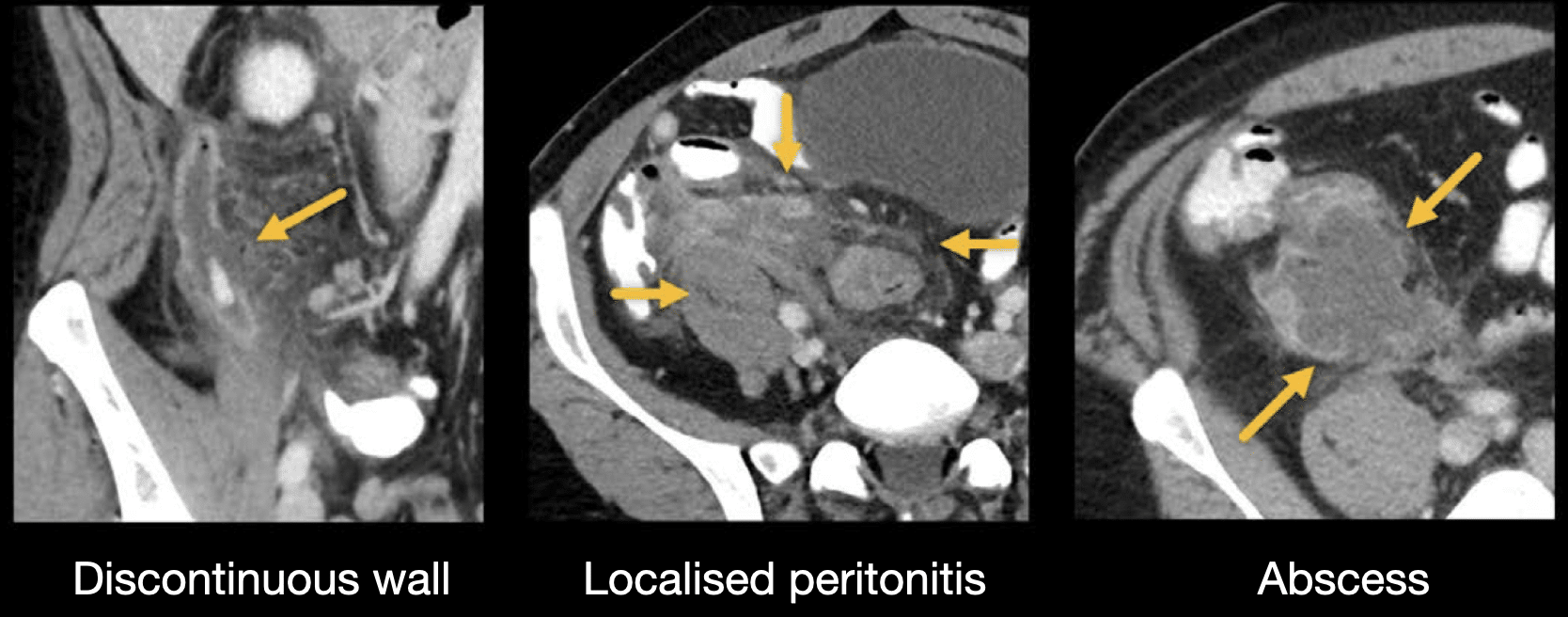
There are three essential imaging findings that will help you characterize complications related to rupture or perforation of the appendix:
- The thin, enhancing wall of the appendix will become discontinuous at the site of perforation
- You will see a large amount of inflammation in the right lower quadrant affecting nearby structures, which can be described as localized peritonitis.
- Abscess formation will occur from the contents that leaked out of the ruptured appendix. An abscess will look like a fluid-density collection with a wall that is variably thick and enhancing (depending on how organized or walled-off the collection is).
Clinical Case 2
Try out an evaluation of the appendix using our online PACS viewer. See which of the key findings mentioned above helps us to make the diagnosis in this case.
- Once again, it is helpful to pull down the coronal images and view them side-by-side with the axial images.
- Scroll to the right lower quadrant and find the dilated, tubular structure behind the cecum with surrounding stranding and inflammation.
- Following the structure down to the cecal base, notice there are two densities in the lumen of the appendix. These were caused by stones or appendicoliths which had obstructed the appendix
- Notice there is a good deal of fluid and small dots of air outside of the appendix. This is indicative of perforation.
- As you look at the inside (medial) wall, notice the gap in the enhancement. The coronal images really highlight this finding, showing a big gap in the wall.
- Also notice the inflammation of the surrounding structures, which is identified by stranding and thickening. This inflammation is due to the content that has spilled out from the perforated appendix, causing localized peritonitis. Over time, this fluid can become organized into a discrete abscess, but that was not the case here.
This is an edited excerpt from the Medmastery course Abdominal CT Pathologies by Michael P. Hartung, MD. Acknowledgement and attribution to Medmastery for providing course transcripts
- Hartung MP. Abdominal CT: Common Pathologies. Medmastery
- Hartung MP. Abdominal CT: Essentials. Medmastery
- Hartung MP. Abdomen CT: Trauma. Medmastery
References
- Connell L, Buttner R. Appendicitis. LITFL
- Cadogan M. Appendicitis – the eponymous examination. LITFL
- Mahmood U. Alvarado score. LITFL
- Top 100 CT scan quiz. LITFL
Radiology Library: Abdominal CT: Imaging important abdominal structures
- Hartung MP. Abdominal CT: acute appendicitis
- Hartung MP. Abdominal CT: diverticulitis
- Hartung MP. Abdominal CT: small bowel obstruction
- Hartung MP. Abdominal CT: closed loop small bowel obstruction
- Hartung MP. Abdominal CT: enteritis and colitis
- Hartung MP. Abdominal CT: peptic ulcer disease
- Hartung MP. Abdominal CT: peptic ulcer perforation
- Hartung MP. Abdominal CT: bowel perforation
Abdominal CT interpretation

Assistant Professor of Abdominal Imaging and Intervention at the University of Wisconsin Madison School of Medicine and Public Health. Interests include resident and medical student education, incorporating the latest technology for teaching radiology. I am also active as a volunteer teleradiologist for hospitals in Peru and Kenya. | Medmastery | Radiopaedia | Website | Twitter | LinkedIn | Scopus

MBChB (hons), BMedSci - University of Edinburgh. Living the good life in emergency medicine down under. Interested in medical imaging and physiology. Love hiking, cycling and the great outdoors.