CT Case 044
A 73-year-old nursing home patient presents with 2 days of abdominal pain and distension, unable to pass a bowel motion or flatus.
He has a grossly distended abdomen, with generalised discomfort on palpation.
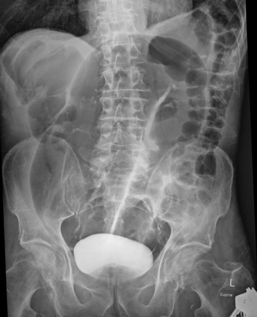
An abdominal X-Ray is performed.

Describe and interpret the Abdominal X-Ray
AXR INTERPRETATION
There are dilated large bowel loops, arising from the pelvis. The dilated bowel is ahaustral (haustra are not seen), unlike in normal loops of bowel.
There is a classical ‘coffee bean sign’. The medial walls of the dilated loops oppose each other, forming a thicker line which gives a coffee bean appearance.
These features are all typical of sigmoid volvulus.
Other signs on x-ray which help in diagnosing sigmoid volvulus are:
- Frimann-Dahl sign: this is seen as three dense lines converging towards site of obstruction in the pelvis. In this patient the point of convergence is obscured to the dense contrast in the bladder
- Absence of rectal gas
Caecal volvulus is a close differential on x-ray imaging and can be differentiated by a few points:
- In caecal volvulus the loops arise from right lower quadrant as opposed to left lower quadrant or pelvis in sigmoid volvulus
- Haustra are seen in caecal volvulus and not in sigmoid volvulus
- Small bowel obstruction is seen in caecal volvulus, whereas colonic obstruction is present in sigmoid volvulus

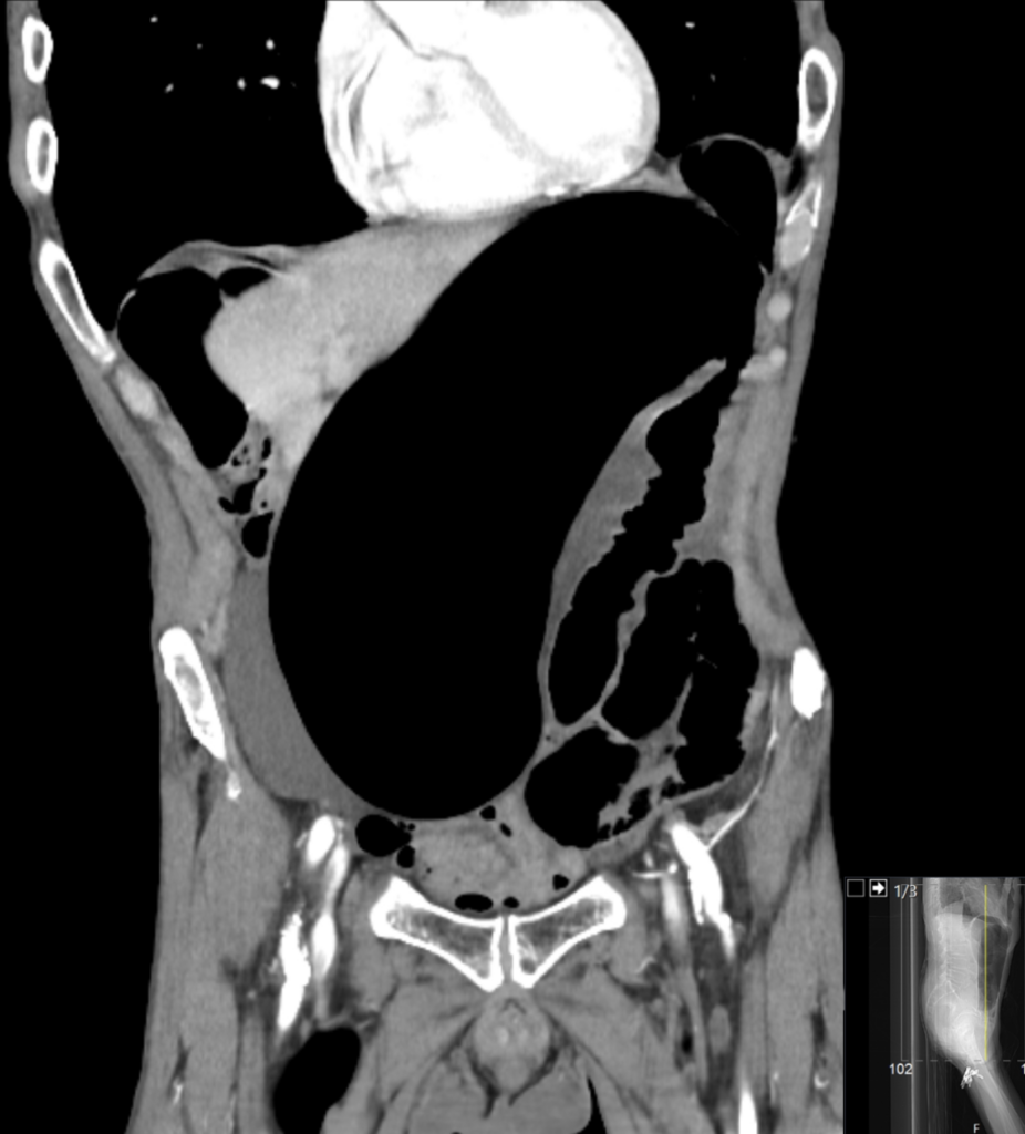
An urgent CT scan is performed to confirm the diagnosis, and assess for associated complications.


Describe and interpret the CT images
CT INTERPRETATION
CT helps in confirming the diagnosis of sigmoid volvulus.
Again, we see distended ahaustral large bowel, and absent rectal gas.
Additionally, on CT scan we see what is called a ‘whirl sign’. This is due to twisting of the sigmoid mesentery.
We look also for complications such as perforation and necrosis, which were fortunately not present in this case.


CLINICAL CORRELATION
A volvulus occurs when a loop of bowel twists around itself and the mesentery that provides its blood supply.
Sigmoid volvulus is the most common type of volvulus of the colon (60%). Other locations are caecal volvulus and transverse colon volvulus.
Patients with chronically distended and redundant colon are at risk; most often this is seen in elderly, bed-bound patients with chronic constipation, it is also seen in those with neurological condition (ie Parkinson’s disease and MS), or in certain African populations with a very high fibre diet combined with an underlying anatomical predisposition.
A volvulus is a closed loop obstruction. This means both the inflow and outflow of the colon are obstructed. The obstructed bowel continues to distend, due to gas forming bacteria trapped inside. If untreated, this will eventually lead to perforation.
Ischaemia gut is the other main complication. As the mesentery is twisted, this compresses the artery and vein, compromising blood supply to the distended bowel.
A sigmoid volvulus is a surgical emergency and requires prompt management to avoid complication.
Most cases are managed with endoscopic detorsion, achieved by sigmoidoscopy and insertion of a flatus tube. Laparoscopy/laparotomy will be required in cases of bowel ischaemia or perforation.
References
- Hartung MP. Abdominal CT: closed loop. LITFL
- Cadogan M. AXR Interpretation. LITFL
- Iqbal S. Sigmoid volvulus. Radiopaedia
TOP 100 CT SERIES
Provisional fellow in emergency radiology, Liverpool hospital, Sydney. Other areas of interest include paediatric and cardiac imaging.
Emergency Medicine Education Fellow at Liverpool Hospital NSW. MBBS (Hons) Monash University. Interests in indigenous health and medical education. When not in the emergency department, can most likely be found running up some mountain training for the next ultramarathon.
Dr Leon Lam FRANZCR MBBS BSci(Med). Clinical Radiologist and Senior Staff Specialist at Liverpool Hospital, Sydney
Sydney-based Emergency Physician (MBBS, FACEM) working at Liverpool Hospital. Passionate about education, trainees and travel. Special interests include radiology, orthopaedics and trauma. Creator of the Sydney Emergency XRay interpretation day (SEXI).


