
![]()
CXR Case 017
67 year old male presents with shortness of breath, cough and significant weight loss. He had a bronchoscpy the following day.
click images to enlarge
Describe and interpret this CXR and subsequent bronchoscopy
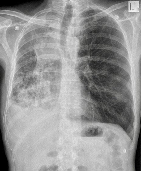
CHEST X-RAY INTERPRETATION
CXR:
There is volume loss in the right hemithorax with collapse of the right upper lobe.
*There is patchy consolidation in the right lower and middle lobes*
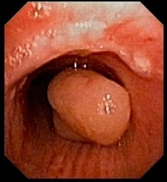
Bronchoscopy:
Bronchoscopy reveals a large obstructing polypoid mass in the right main bronchus.
CLINICAL CORRELATION
The tumour was snared and removed using cautery – its origin was from the right upper lobe.
*The patchy consolidation in the lower and middle lobes is from post-obstructive pneumonia.*
CLINICAL PEARLS
Many endobronchial tumours are benign (e.g. hamartoma), although squamous cell cancer or carcinoid are frequently found.
TOP 150 CXR SERIES
![]()
![]()
![]()
Prof Fraser Brims Curtin Medical School, acute and respiratory medicine specialist, immediate care in sport doc, ex-Royal Navy, academic| Top 100 CXR | Google Scholar | ICIS Course ANZ

Cxr case 017
Is there right side pleural effusion ?
I guess there could be – not a characteristic meniscus laterally, or at least the consolidation around makes it hard to tell. Remember you can hide ~500mls pleural fluid in the costophrenic angle on standard PA film…