
![]()
CXR Case 139
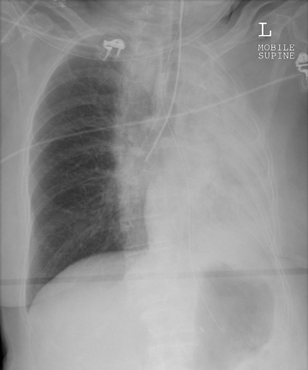
A 53 year old man is intubated in ED for respiratory distress and hypoxaemia.
Describe and interpret this CXR
CHEST X-RAY INTERPRETATION
There is collapse of the left lung with some likely background consolidation. The tip of the endotracheal tube (ETT) is in the right main bronchus.
NG tube is correctly in situ.
Right lung parenchyma and pleura look clear.
CLINICAL CORRELATION
The white out of the left side is collapse because the right heart border and trachea are markedly displaced to the left – indicating volume loss on the left.
It is not clear if the pneumonia is responsible for the collapse, the ETT is in too far, which may be responsible, or both.
CLINICAL PEARLS
Pull the tube back, repeat the CXR and allow some good old positive pressure to do the rest.
More iatrogenic mishaps at Top 10 X-Ray foreign bodies…
TOP 150 CXR SERIES
Prof Fraser Brims Curtin Medical School, acute and respiratory medicine specialist, immediate care in sport doc, ex-Royal Navy, academic| Top 100 CXR | Google Scholar | ICIS Course ANZ