
![]()
ECG Case 048
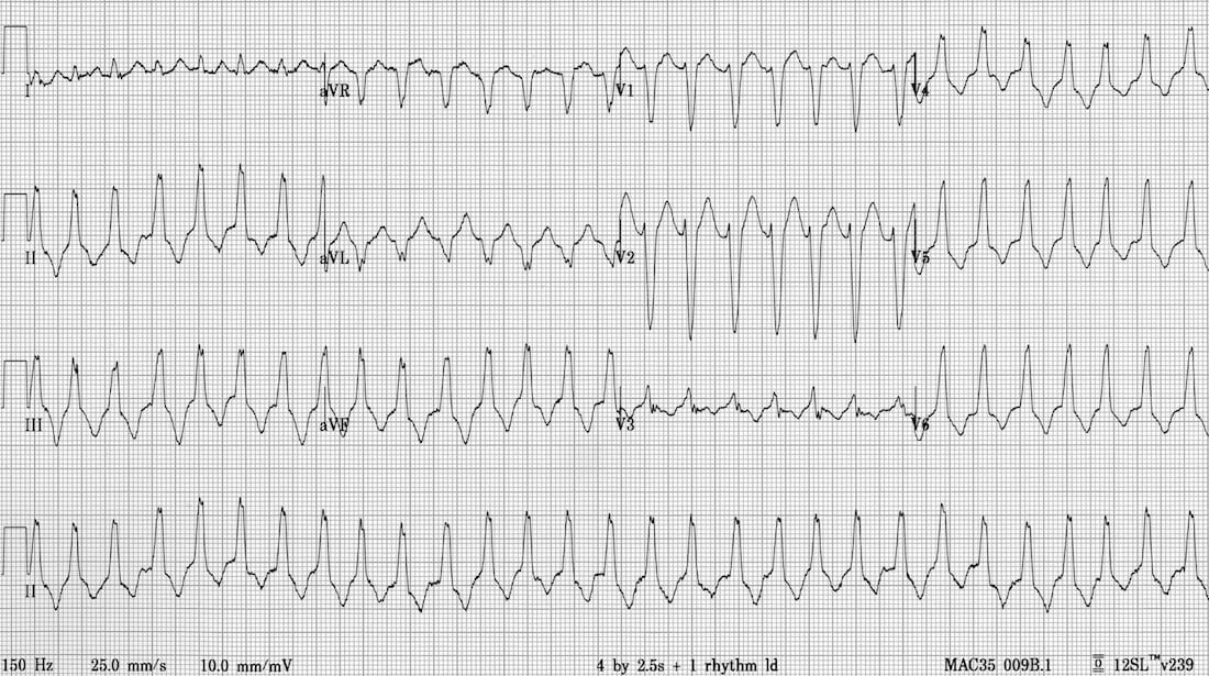
30-year old female presenting with sudden onset of palpitations. Normally well. Describe the ECG.
Describe and interpret this ECG
ECG ANSWER and INTERPRETATION
On first glance this would appear to be SVT with LBBB as there is:
- Regular broad-complex tachycardia.
- No atrial activity seen.
- Typical LBBB morphology in aVR, V1 and V6.
- No obvious diagnostic features for VT — compare this with ECG 047.
However, there is one feature here that is unusual for LBBB, can you spot it?
Reveal answer
There is an rightward / inferior axis (around +90 degree)., which is atypical for Left Bundle branch block. LBBB normally has a leftward axis.
This combination of…
- Broad complex tachycardia with typical LBBB morphology.
- Inferior axis (+90 degrees).
… is suggestive of a specific type of VT known as right ventricular outflow-tract tachycardia (RVOT).
RVOT is a relatively common form of right ventricular VT, occurring in two main groups:
- Patients with structurally normal hearts (= 70% of idiopathic VT).
- Patients with arrhythmogenic right ventricular cardiomyopathy.
It may be very difficult to differentiate RVOT from SVT with LBBB.
[NB. Left bundle branch block morphology simply indicates that the heart is depolarising from right to left. Hence, similar QRS patterns are seen with LBBB, RVOT and RV-pacing]
CLINICAL PEARLS
Tips for Spotting RVOT
- Suspect RVOT when you see LBBB morphology + inferior axis.
- Record a long rhythm strip looking for fusion and capture beats.
I have diagnosed this only a couple of times in the past. Each time I had to stand by the monitor with my finger on the “print” button waiting for a fusion or capture beat to appear before anyone would believe me!
References
Further Reading
- Wiesbauer F, Kühn P. ECG Mastery: Yellow Belt online course. Understand ECG basics. Medmastery
- Wiesbauer F, Kühn P. ECG Mastery: Blue Belt online course: Become an ECG expert. Medmastery
- Kühn P, Houghton A. ECG Mastery: Black Belt Workshop. Advanced ECG interpretation. Medmastery
- Rawshani A. Clinical ECG Interpretation ECG Waves
- Smith SW. Dr Smith’s ECG blog.
- Wiesbauer F. Little Black Book of ECG Secrets. Medmastery PDF
TOP 100 ECG Series
Emergency Physician in Prehospital and Retrieval Medicine in Sydney, Australia. He has a passion for ECG interpretation and medical education | ECG Library |

Very nice ECG. I believe there is also AV dissociation apparent upon closer inspection (best seen V1, V4 and the rhythm strip), which pushed me in the direction of VT. I thought that would be a nice addition in the line of the previous two cases.
Thanks for these ECG’s. They are helping me in my 12 lead training.