
![]()
Eyes Wide Split
aka Ophthalmology Befuddler 013
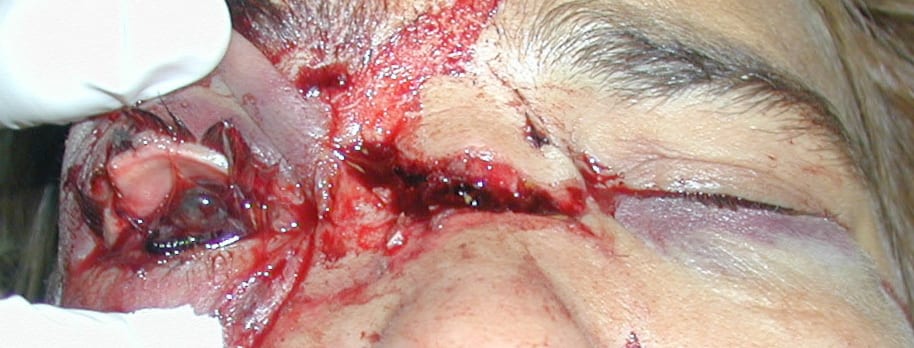
A 38 year-old truck driver had an argument with his wife after they had been out for a few drinks. Unfortunately, the altercation got out of hand and she threw a brick at his face and then punched him in the face.
Even more unfortunate was the fact that she was wearing the giant ring he presented to her on their tenth wedding anniversary — one of its sharp edges caught his right eye.
Questions
Q1. What are the different types of penetrating eye trauma?
Answer and interpretation
The major types of penetrating eye injury are:
- eye lid lacerations
- corneal lacerations
- scleral lacerations
- perforating trauma (+/- an exit wound) including occult foreign body penetration (e.g. when metal strikes metal)
There may also be associated injuries to:
- intraocular structures — e.g. lens, iris, retina
- extraocular structures — e.g. lids, extra-ocular muscles, orbital bones, optic nerve and brain
Q2. What features on history should be assessed?
Answer and interpretation
History:
- symptoms — visual disturbance or loss of vision, pain at rest or on movement, and diplopia.
- mechanism of injury — any suspicion of penetrating eye trauma requires prompt assessment so that urgent ophthalmology referral can be made.
- Use of eye protection
- type of projectile and velocity — small high-velocity projectiles are at higher risk of penetrating injury.
- history of previous trauma or surgery that may compromise the structural integrity of the eye.
in addition to the usual AMPLE history for trauma.
Q3. What features on examination should be assessed?
Answer and interpretation
Examination — if penetrating injury is possible, but not obvious, carefully assess for:
- pupil — a teardrop-shaped pupil may be caused by iris prolapse through a corneal laceration.
- slit lamp — look for defects in the cornea or sclera or distortion of the anterior chamber structures (e.g. a shallow anterior chamber with a self-sealing corneal laceration), hyphaema. Check under the eyelids for a concealed laceration/ rupture.
- visual acuity — usually decreased
- red reflex — may be abnormal
- fundoscopy — look for foreign bodies and retinal injury
If penetrating injury is obvious, only a cursory examination is needed — make the referral!
Q4. What is the management of penetrating trauma to the eye?
Answer and interpretation
Urgent referral to an ophthalmologist is indicated. CT scan of the orbit may be performed following discussion with an ophthalmologist to check for ocular or orbital foreign bodies.
Management Involves:
- The patient is kept nil by mouth, with strict bed rest.
- Supportive care including analgesia and antiemetics as required.
- Apply an eye shield (not a pad) to protect the eye, but avoid applying pressure that will increase intraocular pressure leading to extrusion of ocular contents.
- Start broad spectrum IV antibiotics (see Q9). Do not apply any topical agents if there is a penetrating eye injury as the preservative is toxic to ocular contents.
- Tetanus immunisation as required
Q5. What are the complications of penetrating eye injury?
Answer and interpretation
- permanent loss of vision — enucleation may be necessary
- corneal ulcers — may lead to delayed perforation.
- infection — e.g. endophthalmitis
- sympathetic ophthalmia — enucleation of the severely traumatised eye should be performed initially or within 1-2 weeks to prevent this in the severely damaged eye
Q6. What is Seidel test?
Answer and interpretation
Seidel test is used to detect aqueous humor leaking from a corneal wound. (see: Something in my Eye Doc)
Fluorescein dye is applied to the region of the suspected laceration. The test is positive when a stream of fluorescent dye emanating from the site is visualised on slit-lamp examination.
A negative test does not rule out a full thickness corneal laceration.
Seidel positive Test (RootAtlas)
Q7. How are conjunctival lacerations managed?
Answer and interpretation
- Superficial wounds less than about 1-1.5cm long generally heal spontaneously. If the conjunctiva is rolled back on itself it may need to be realigned.
- Deeper and more extensive lesions require sutures (e.g. 9-0 absorbable), and are usually repaired by an ophthalmologist.
- Large lacerations are seen for follow up in a week, small lacerations can be reviewed as needed.
Q8. What is sympathetic ophthalmia?
Answer and interpretation
Inflammation of the uninjured eye occurring weeks to months after the initial insult to the injured eye.
- pathophysiology — an autoimmune response to the normally sequestered uveal tissues of the injured eye becoming exposed with injury.
- clinical manifestations — pain, photophobia, and decreased visual acuity.
- management — ophthalmology referral for treatment with steroids and immunosuppressants. Symptoms may be reduced by enucleation of the blind injured eye even after sympathetic ophthalmia has developed.
Q9. What is endophthalmitis?
Answer and interpretation
Endophthalmitis is inflammation, often due to infection, involving all the deep structures of the eye.
- Pathophysiology — complication of blunt globe rupture, penetrating eye injury, foreign bodies, and ocular surgery. Causative organisms are usually Staphylococcus, Streptococcus, and GNBs (e.g. Bacillus).
- Clinical manifestations — pain and visual loss; chemosis, and hyperemia of the conjunctiva, and the infected chambers are hazy or opaque.
- Management — topical, intraocular and systemic antibiotics
The Australian Therapeutic Guidelines suggests the following, unless alternative specialist advice is given:
- ciprofloxacin 750 mg (child: 20 mg/kg up to 750 mg) orally, as a single dose
- vancomycin 25 mg/kg up to 1.5 g (child less than 12 years: 30 mg/kg up to 1.5 g) IV, as a single dose
References
- Ehlers JP, Shah CP, Fenton GL, Hoskins EN. The Wills Eye Manual: Office and Emergency Room Diagnosis and Treatment of Eye Disease Lippincott Williams & Wilkins
- NSW Statewide Opthalmology Service. Eye Emergency Manual — An illustrated Guide. [Free PDF]
OPHTHALMOLOGY BEFUDDLER
Chris is an Intensivist and ECMO specialist at The Alfred ICU, where he is Deputy Director (Education). He is a Clinical Adjunct Associate Professor at Monash University, the Lead for the Clinician Educator Incubator programme, and a CICM First Part Examiner.
He is an internationally recognised Clinician Educator with a passion for helping clinicians learn and for improving the clinical performance of individuals and collectives. He was one of the founders of the FOAM movement (Free Open-Access Medical education) has been recognised for his contributions to education with awards from ANZICS, ANZAHPE, and ACEM.
His one great achievement is being the father of three amazing children.
On Bluesky, he is @precordialthump.bsky.social and on the site that Elon has screwed up, he is @precordialthump.
| INTENSIVE | RAGE | Resuscitology | SMACC