


George Perkins
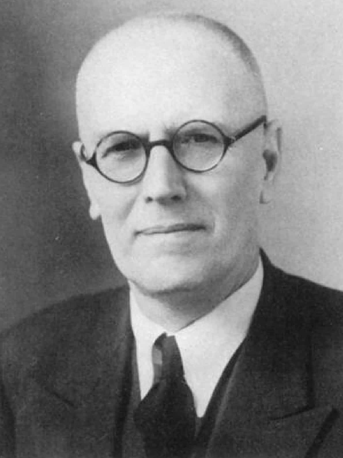
George Perkins (1892-1979) was an English orthopaedic surgeon
Perkins was an influential surgical teacher at St Thomas’ Hospital, London. He served in both world wars, helped build the St Thomas’ orthopaedic department with Rowley Bristow, and became its head before being appointed the hospital’s first Professor of Surgery in 1948. Perkins was also instrumental in creating the British Volume of the Journal of Bone and Joint Surgery
He is remembered for Perkins line, used in radiographic assessment of congenital hip dislocation, and Perkins traction, a functional method of femoral fracture treatment based on skeletal traction with early active joint movement. His rejection of prolonged immobilisation in favour of function and mobilisation was considered radical by many contemporaries.
Biographical Timeline
- Born on September 22, 1892 in Staines, Middlesex.
- 1914 – Enlisted in the Army at the outbreak of the First World War but was sent back to complete his medical studies.
- 1916 – Qualified MB, BCh Oxford (Hertford College) and immediately joined the Royal Army Medical Corps (RAMC). Posted to East Africa as medical officer to the 3rd King’s African Rifles, initially with the rank of Captain.
- 1917–1918 – Awarded the Military Cross during the East African Campaign. He was subsequently captured by German forces and escaped.
- 1919 – Demobilised and returned to St Thomas’ as a house surgeon. He worked under Sir Max Page and Percy Sargent. Later, worked at the Military Orthopaedic Hospital, Shepherd’s Bush with Sir Robert Jones (1857–1933) and Naughton Dunn (1884–1939).
- 1921 – Obtained FRCS and MCh and won the first Robert Jones Prize. Elected honorary assistant surgeon to the Royal National Orthopaedic Hospital
- 1924 – Became chief assistant to Rowley Bristow in the newly established Orthopaedic Department at St Thomas’. The collaboration helped develop the department into a major orthopaedic centre.
- 1928 – Published “Signs by which to diagnose congenital dislocation of the hip”, describing radiographic assessment that included a vertical line through the lateral acetabular margin. This subsequently became known as Perkins line.
- 1929 – Appointed assistant orthopaedic surgeon at St Thomas’ Hospital. T
- 1939 – Recalled to the RAMC as a surgical specialist with the rank of Major at the outbreak of the Second World War and served with a casualty clearing station in France.
- 1940–1941 – Invalided home because of serious illness. During his convalescence he began writing on fracture treatment.
- 1944 – Joined the staff of Queen Mary’s Hospital, Roehampton, working in amputation and chronic osteomyelitis.
- 1946 – Succeeded Rowley Bristow as head of the Orthopaedic Department at St Thomas’ and became President of the British Orthopaedic Association. As BOA President, strongly promoted establishment of an independent British voice for the Journal of Bone and Joint Surgery.
- 1948 – Appointed the first Professor of Surgery at St Thomas’ Hospital / University of London.
- 1948–1952 – Chaired the British Editorial Board of the Journal of Bone and Joint Surgery after Sir Harry Platt.
- 1940s–1950s – Developed an unconventional approach to fracture management, emphasising traction, function and early active movement rather than prolonged rigid plaster immobilisation.
- 1954–1955 – Published Foundations of Surgery and stepped down from the professorial unit.
- 1961 – Published the major textbook Orthopaedics.
- 1970 – Published The Ruminations of an Orthopaedic Surgeon, reflecting his provocative and questioning approach to surgical practice.
- Died on October 22, 1979 aged 87.
Medical Eponyms
Perkins line (1928)
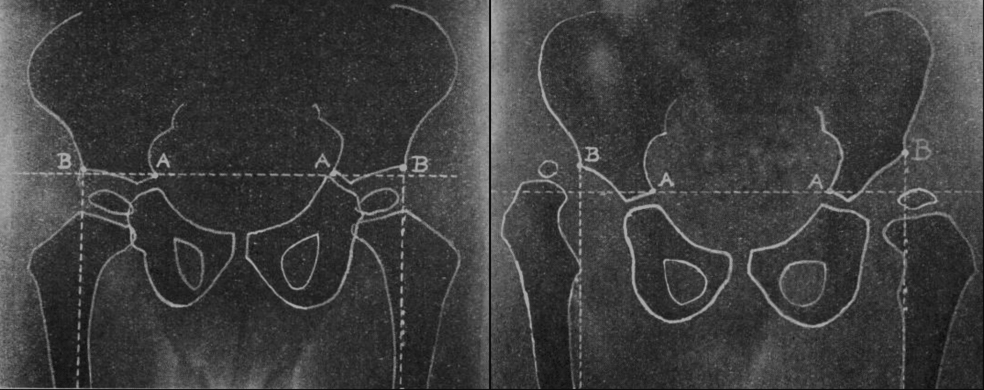
Perkins line (Perkins’ line; Perkin line) is the vertical radiographic reference line drawn perpendicular to Hilgenreiner line through the lateral acetabular margin on an AP paediatric pelvis radiograph. Together with Hilgenreiner line it creates four quadrants. The normal proximal femoral epiphysis should lie in the inferomedial quadrant.
1928 – Perkins described a two-line construction in Signs by which to diagnose congenital dislocation of the hip. A horizontal line through the Y-shaped triradiate cartilages and a perpendicular dropped from the prominent lateral acetabular margin. He used these lines to demonstrate superior and lateral displacement of the proximal femur in congenital hip dislocation.
These two lines will show whether the head of the femur is further up or further out than it should be
Perkins traction (1958)
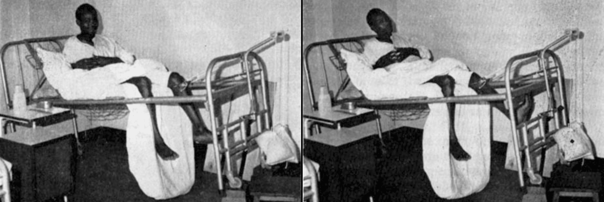
Perkins traction is a method of skeletal traction for fractures of the femur, particularly shaft, distal and comminuted fractures. It is designed to maintain alignment while permitting early active movement of the knee.
The technique uses skeletal traction through a tibial pin, elevation of the foot of the bed and counter-traction from body weight. The lower part of the bed is opened or lowered so that the patient can begin repeated active knee flexion and extension while the fracture remains in traction.
Commemorative eponyms / legacy
Perkins Lecture
The Perkins Lecture at St Thomas’ Hospital was established in his honour. The inaugural lecture was delivered by Maurice E. Müller (1918–2009) on rigid internal fixation, a technique Perkins supported because effective fixation allowed joints to be mobilised rather than externally immobilised.
George Perkins Ward
St Thomas’ Hospital named a ward after George Perkins following his death.
Key Medical Contributions
Functional fracture treatment
Perkins challenged the prevailing doctrine of prolonged rigid immobilisation and emphasised “treating the patient and not the X-ray”
- traction to control alignment;
- active movement as early as possible;
- preservation of joint function;
- clinical rather than exclusively radiographic assessment of union.
His contemporaries regarded this approach as radically different from the extensive splintage promoted by Böhler and Watson-Jones.
Major Publications
- Perkins G. Signs by which to diagnose congenital dislocation of the hip. Lancet 1928: 648-650
- Perkins G. Rest and movement. J Bone Joint Surg Br. 1953 Nov;35-B(4):521-39.
- Perkins G. Foundations of Surgery. 1954
- Perkins G. Fractures and Dislocations. 1958
- Perkins G. Orthopaedics. 1961
- Perkins G. The ruminations of an orthopaedic surgeon. 1970
- Perkins G. Signs by which to diagnose congenital dislocation of the hip. 1928. Clin Orthop Relat Res. 1992 Jan;(274):3-5
Perkins reportedly completed the publications section of his own CV with:
“None of any importance except to myself.”
References
Biography
- Apley AG. George Perkins. J Bone Joint Surg Br. 1973 Feb;55(1):4-6.
- Professor George Perkins. The Daily Telegraph Thursday, November 22, 1979
- G Perkins. Br Med J 1980;280:58
- In memoriam George Perkins, 1892-1979. J Bone Joint Surg Br. 1980 May;62-B(2):248-50.
- Mostofi SB. George Perkins (1892–1979). In: Who’s Who in Orthopedics. Springer, London 2005: 265-267
- Perkins, George (1892-1980). Plarr’s Lives of the Fellows
Eponymous terms
- Bewes PC. Fractures of the femur in a tropical context: a re-evaluation of Perkins’ traction. Trop Doct. 1974 Apr;4(2):64-8.
- Gosselin R, Lavaly D. Perkins traction for adult femoral shaft fractures: a report on 53 patients in Sierra Leone. Int Orthop. 2007 Oct;31(5):697-702.
Eponym
the person behind the name

MB BAO BCh Trinity College Dublin, MSc Clinical Data Analytics University of Galway. Junior doctor in Perth, doctoral researcher in MRI radiomics. Working towards radiology training.

BA MA (Oxon) MBChB (Edin) FACEM FFSEM. Emergency physician, Sir Charles Gairdner Hospital. Passion for rugby, medical history, medical education, and asynchronous learning #FOAMed evangelist. Editor-in-Chief and Webmaster of Life in the Fast lane, LITFL | On Call 4e| Eponyms | Books | Horology |