


Perkins line
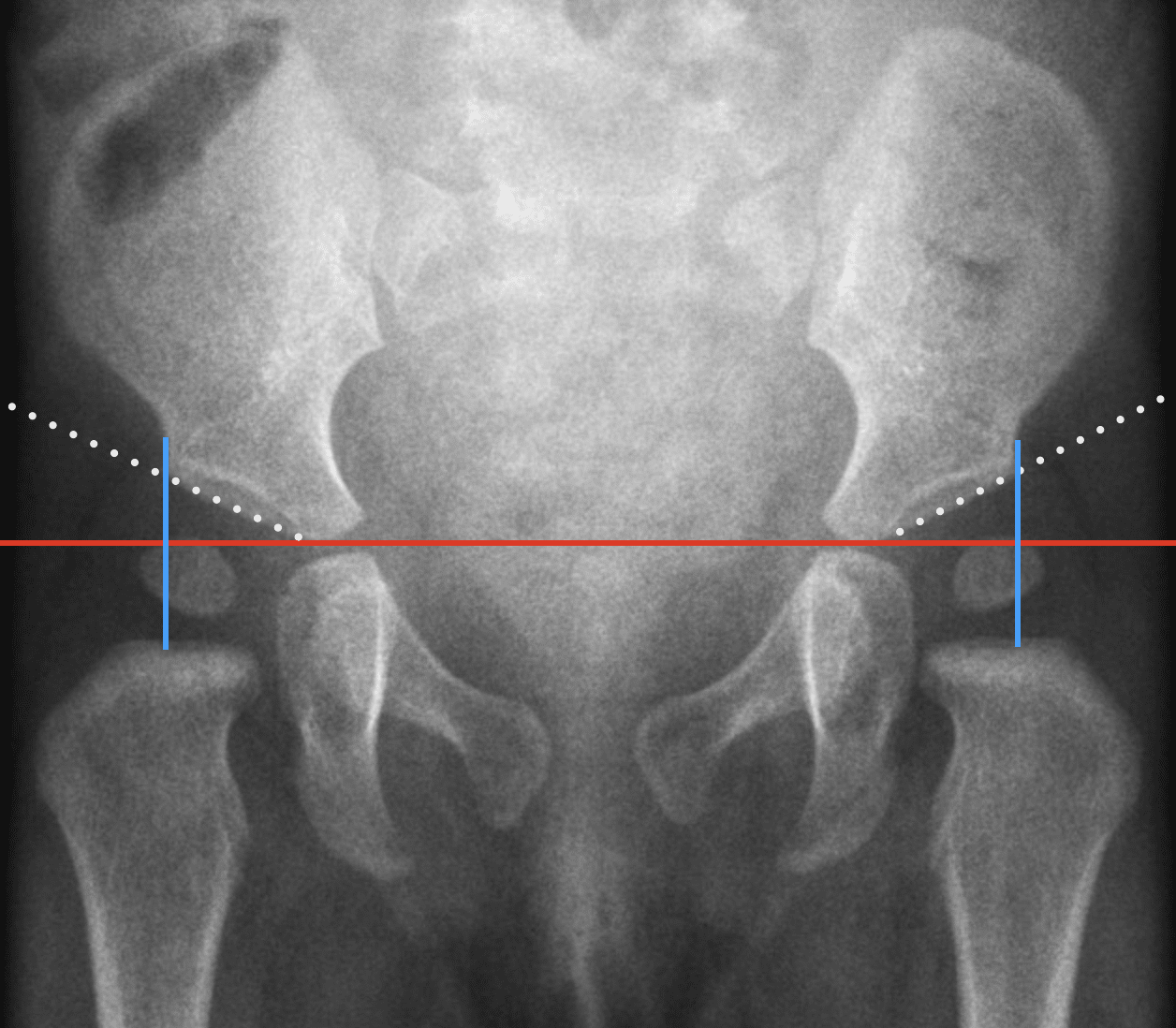
Perkins line (Perkin line; Perkins’ line, Ombrédanne-Perkins line) is a vertical radiographic reference line drawn perpendicular to Hilgenreiner line through the lateral acetabular margin on an AP radiograph of the paediatric pelvis. Together, the two lines divide each hip into quadrants and are used in assessment of developmental dysplasia of the hip (DDH).
In the normally located hip, the ossified femoral-head epiphysis should lie below Hilgenreiner line and medial to Perkin line, within the inferomedial quadrant. When the femoral head is not yet ossified, the position of the proximal femur relative to these landmarks remains useful. Modern descriptions specifically define Perkin line as perpendicular to Hilgenreiner line at the lateral acetabular roof.
History of the Perkins line
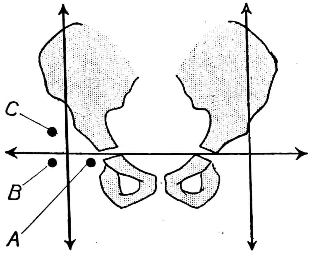
1923 – Louis Ombrédanne provided the first known description of a vertical acetabular reference. Ombrédanne drew a horizontal line through the bilateral acetabular-floor shadows and a vertical tangent to the lateral border of the acetabular roof. The two references localised the femoral-head ossific nucleus: normal inferomedial position (A), early lateral displacement (B), and established superolateral displacement (C).
1925 – Heinrich Hilgenreiner (1870–1953) described a horizontal baseline connecting the two Y-shaped (triradiate) cartilages as part of a wider measurement system for congenital hip dislocation. He extended this line laterally and used perpendicular measurements from the proximal femora to quantify superior displacement, lateral displacement and acetabular inclination.
My procedure is to draw a horizontal line connecting the two Y-shaped cartilages.
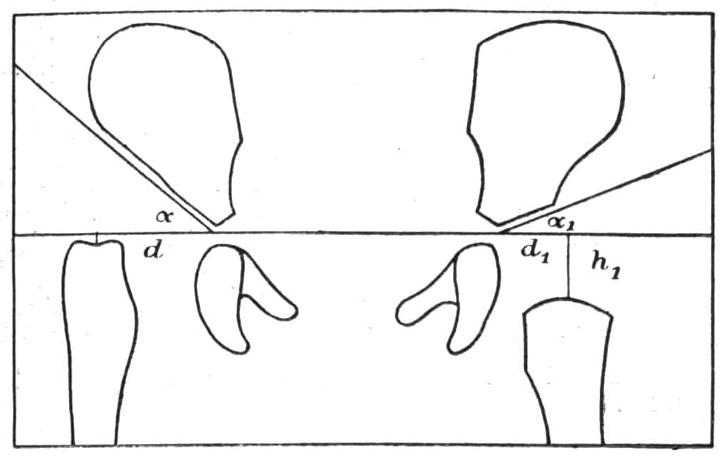
Importantly, Hilgenreiner’s perpendiculars arose from the proximal femora. They were therefore not the modern Perkin lines.
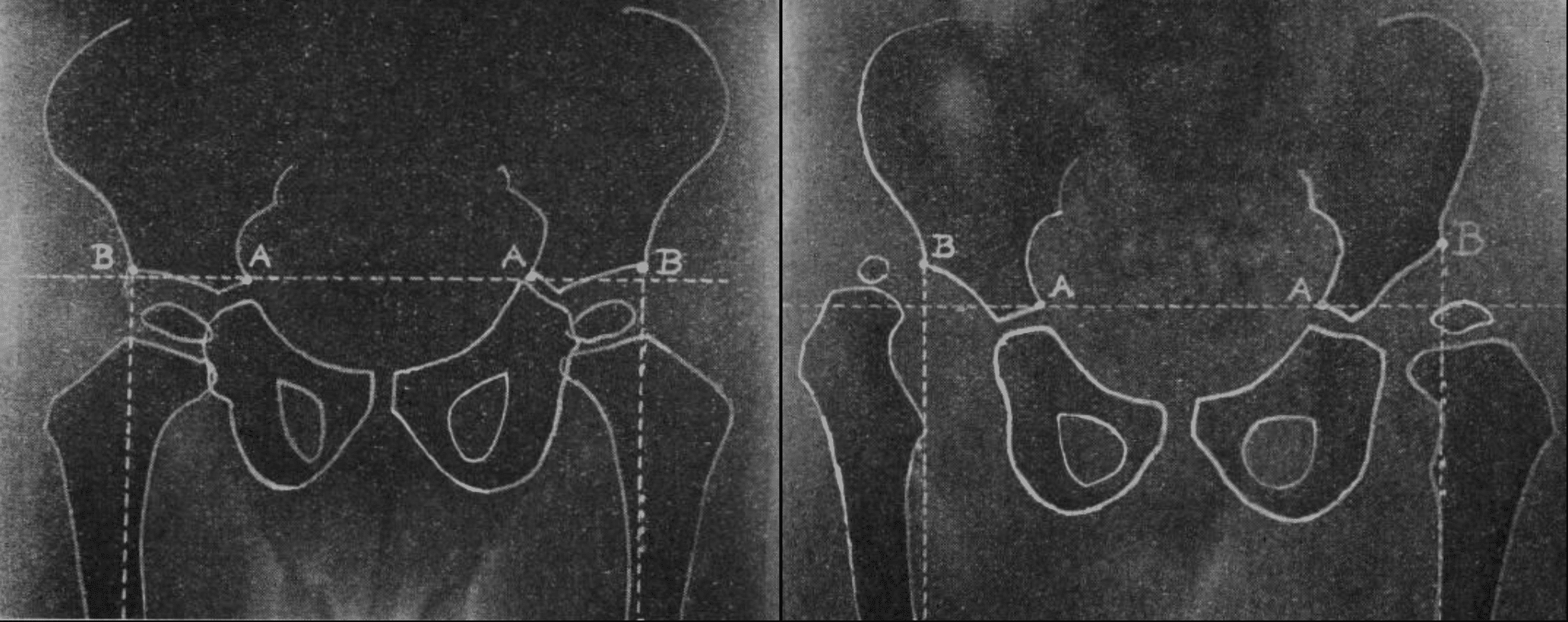
1928 – George Perkins (1892–1979) published Signs by which to diagnose congenital dislocation of the hip in The Lancet. His aim was to provide a simple radiographic method that could show whether the femoral head was displaced upwards or outwards. Perkins first drew his own transverse reference:
Draw a horizontal line joining A to A; A is the innermost part of the ilium at the Y-shaped cartilage of the acetabulum.
He then drew a perpendicular through the lateral pelvic landmark B. The femoral-head ossification centre should lie below the horizontal line and medial to the perpendicular. Superior or lateral displacement suggested congenital dislocation
These two lines will show whether the head of the femur is further up or further out than it should be
1932 – Vittorio Putti (1880–1940) published Analisi della triade radiosintomatica degli stati di prelussazione [Analysis of the radiographic symptom triad of preluxation]. Reimers and Bialik cite Putti’s line as synonymous with Perkins line. The original 1932 article has not yet been directly reviewed, so the precise construction remains to be confirmed.
1951 – Harold E. Martin published Geometrical-anatomical factors and their significance in the early x-ray diagnosis of hip-joint disease in children. Martin described a horizontal Y-line through the cotyloid notches and a V-line dropped vertically from the lateral edge of the superior acetabular lip. Normally the femoral-head epiphysis lay below the Y-line and medial to the V-line. However with subluxation it could lie lateral to the V-line while remaining below the Y-line. With complete dislocation it lay superior and lateral to their intersection.
Martin introduced the construction as part of “Hilgenreiner’s lines”, referring simply to the horizontal Y-line and vertical V-line. The term Martin’s V-line therefore appears to be a later attribution
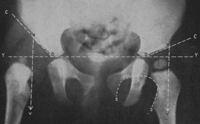
1980 – Jørgen Reimers incorporated Perkin lines perpendicular to Hilgenreiner line into his radiographic assessment of hip migration in children with cerebral palsy. The resulting migration percentage quantified the proportion of the femoral head lying lateral to the acetabular boundary. This embedded the Hilgenreiner–Perkin construction in paediatric hip assessment.
Ombrédanne predates both Hilgenreiner and Perkins for use of a vertical tangent at the lateral acetabular roof to assess lateral femoral-head displacement. Perkins independently described a closely corresponding construction in 1928. The modern eponym “Perkins line” became attached to the vertical reference used with Hilgenreiner line.
Associated Persons
- Louis Ombrédanne (1871-1956)
- Heinrich Hilgenreiner (1870-1953) and the Hilgenreiner line
- Edward Warren Hine Shenton (1872–1955) and the Shenton line
- George Perkins (1892-1979) and the Perkins line
- Louis Ombrédanne (1871-1956)
- Dietrich Tönnis (1927-2010) and the Tönnis angle
- Jørgen Reimers (1931-2008)
Alternative names
- Perkin line, Perkin’s line, Perkins’ line
- Ombrédanne-Perkins line, Ombrédanne line, Ombrédanne–Perkins vertical
Historical synonyms / related terms
- Putti line
- Martin V-line
Eponymythology
The modern Perkins line carries Perkins name, but the vertical acetabular reference was not first described by Perkins. Louis Ombrédanne published a vertical tangent to the lateral acetabular roof in 1923, five years before the Perkins 1928 paper. Ombrédanne combined it with a horizontal pelvic reference to classify the femoral-head ossific nucleus as inferomedial, lateral or superolateral.
Perkins independently described a closely related horizontal–vertical construction in 1928. His method became integrated with Hilgenreiner’s 1925 horizontal baseline, producing the familiar Hilgenreiner–Perkin quadrant system used today.
Later literature further complicated the nomenclature by treating Ombrédanne line, Perkins line, Putti line and Martin V-line as synonyms. These names describe closely related vertical acetabular references, but their original descriptions were not historically simultaneous or necessarily identical.
Hilgenreiner’s vertical lines are not Perkin lines.
Hilgenreiner starts with the femur. He drops a perpendicular from the centre of the proximal femur onto his horizontal Y-cartilage baseline. The position of that perpendicular therefore changes when the femur moves. He then derives h and d from that construction.
Perkins starts his vertical from the pelvis. Point B is a fixed pelvic landmark — explicitly the anterior inferior iliac spine / prominent outer lower iliac angle on the radiograph — and he drops a vertical from B to intersect his A–A transverse line.
The modern Hilgenreiner–Perkin construction represents a later synthesis of two closely related but not identical historical constructions.
References
Historical references
- Hilgenreiner H. Zur Frühdiagnose und Frühbehandlung der angeborenen Hüftgelenkverrenkung. Medizinische Klinik 1925; 21: 1385-1388, 1425-1429
- Perkins G. Signs by which to diagnose congenital dislocation of the hip. Lancet 1928: 648-650
- Perkins G. Signs by which to diagnose congenital dislocation of the hip. 1928. Clin Orthop Relat Res. 1992 Jan;(274):3-5
- Ombrédanne L. Précis clinique et opératoire de chirurgie infantile. 1932: 1113
- Putti V. Analisi della triade radiosintornatica degli staff di prelussazione. La Chirurgia degli organi di movimento 1932;17(5):453–459.
- Martin HE. Geometrical-anatomical factors and their significance in the early x-ray diagnosis of hip-joint disease in children. Radiology. 1951 Jun;56(6):842-9.
- Reimers J, Bialik V. Influence of femoral rotation on the radiological coverage of the femoral head in children. Pediatr Radiol. 1981;10(4):215-8.
Eponymous term review
- Campbell SE. Radiography of the hip: lines, signs, and patterns of disease. Semin Roentgenol. 2005 Jul;40(3):290-319.
- Yeap PM, Budak MJ. The pelvic radiograph: lines, arcs and stripes. Singapore Med J. 2021 Jul;62(7):333-340.
- Bi AS, Carter C, Price AE, Litrenta J, Karamitopoulos M, Castañeda PG. A history of eponym usage in hip and pelvis radiography part 1: the paediatric hip. Hip Int. 2023 Mar;33(2):136-143.
- Bi AS, Ryan MK, Castañeda PG, Youm T. A history of eponym usage in hip and pelvis radiography part 2: the adult hip. Hip Int. 2023 Mar;33(2):144-151
- Kyle E, Cadogan M. Hip and pelvic lines and arcs. LITFL
eponymictionary
the names behind the name

MB BAO BCh Trinity College Dublin, MSc Clinical Data Analytics University of Galway. Junior doctor in Perth, doctoral researcher in MRI radiomics. Working towards radiology training.

BA MA (Oxon) MBChB (Edin) FACEM FFSEM. Emergency physician, Sir Charles Gairdner Hospital. Passion for rugby, medical history, medical education, and asynchronous learning #FOAMed evangelist. Editor-in-Chief and Webmaster of Life in the Fast lane, LITFL | On Call 4e| Eponyms | Books | Horology |