


Hilgenreiner line
Hilgenreiner line is a horizontal radiographic reference line drawn through the bilateral triradiate cartilages on an AP pelvis radiograph in infants and young children. It is used in the assessment of developmental dysplasia of the hip (DDH).
The line provides a horizontal baseline for measurement of the acetabular index and is commonly used with the perpendicular Perkins line to assess the position of the proximal femur. In the normally located hip, the femoral head or proximal femoral epiphysis should lie below Hilgenreiner line and medial to Perkins line, within the inferomedial quadrant. Shenton line provides an additional assessment of hip alignment.
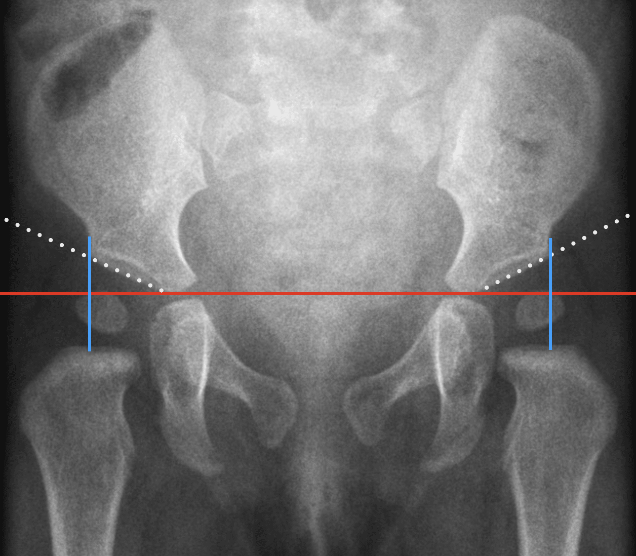
The Hilgenreiner line is commonly interpreted with Perkins line, Shenton line and Reimers migration percentage in assessment of the paediatric hip. See Pelvic radiographic lines and landmarks for their anatomical relationship.
Hilgenreiner line forms the horizontal baseline for the Tönnis angle (acetabular index), used to quantify acetabular roof inclination in the radiographic assessment of developmental hip dysplasia.
History
1925 – Heinrich Hilgenreiner (1870–1953) published Zur Frühdiagnose und Frühbehandlung der angeborenen Hüftgelenkverrenkung in two consecutive issues of Medizinische Klinik. He described a radiographic method capable of identifying congenital hip displacement in infancy, before ossification of the femoral head made conventional landmarks visible.
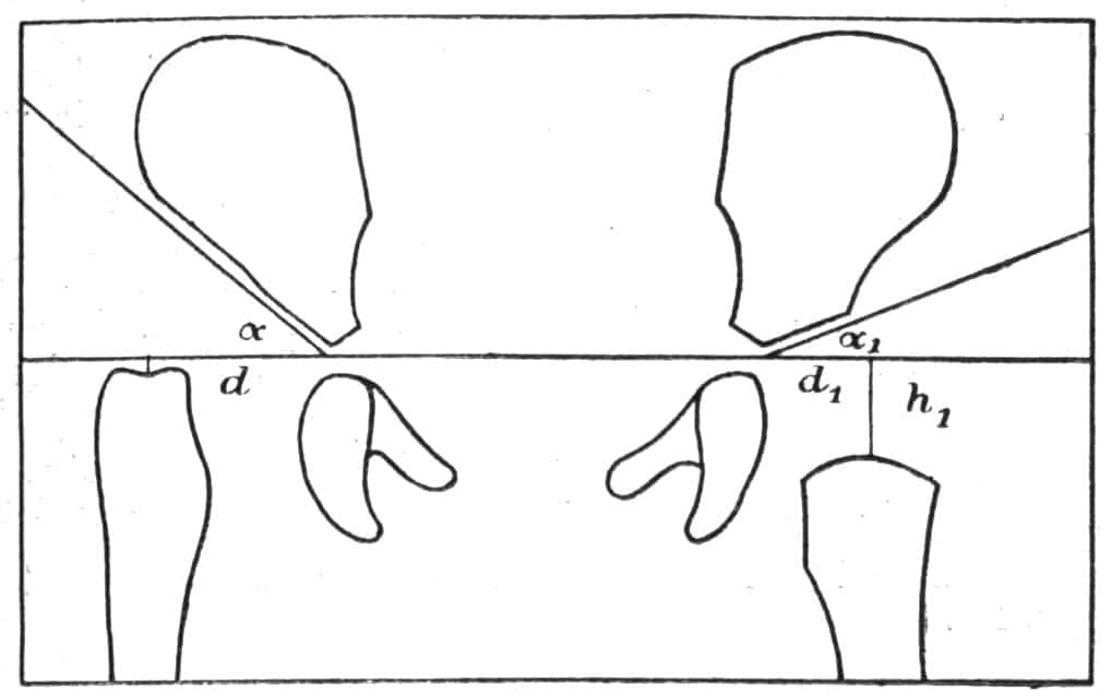
Hilgenreiner used the line connecting the two triradiate (Y-) cartilages as a horizontal baseline. From the centre of the proximal femur he dropped a perpendicular to this baseline and measured h, representing vertical femoral position, and d, representing lateral displacement from the acetabulum. He also measured the acetabular roof angle (α) relative to the baseline.
My procedure is to draw a horizontal line connecting the two Y-shaped cartilages
In the normal infant pelvis he reported an acetabular angle of approximately 20°, h ≈1 cm, and d ≈1–1.5 cm. With congenital dislocation, the acetabular angle and lateral distance increased while h decreased, sometimes reaching zero or becoming negative.
Hilgenreiner’s horizontal baseline became paired with the perpendicular Perkins line, drawn through the lateral acetabular margin, creating the familiar four-quadrant assessment of the paediatric hip.
1986 – An English translation of Hilgenreiner’s 1925 paper was published as Hilgenreiner on congenital hip dislocation in the Journal of Pediatric Orthopaedics, making the original description more accessible to English-language readers.
Modern use: Hilgenreiner line remains a standard reference in paediatric pelvic radiography, forming the baseline for the acetabular index and being used with Perkins and Shenton lines in assessment of hip position and DDH.
Associated Persons
- Heinrich Hilgenreiner (1870-1953) and the Hilgenreiner line
- Edward Warren Hine Shenton (1872–1955) and the Shenton line
- George Perkins (1892-1979) and the Perkins line
- Dietrich Tönnis (1927-2010) and the Tönnis angle
Alternative names
- Hilgenreiner’ Hilfslinien, Hilgenreiner Skiagram,
- Hilgenreiner Beckenskiagram, Hilgenreiner MessungName
Eponymythology
Hilgenreiner did not originally describe a single isolated line. His 1925 method was a radiographic system based on a horizontal line through the triradiate cartilages, with measurements of proximal femoral height (h), lateral displacement (d) and acetabular inclination (α).
Modern measurements retain the horizontal baseline as Hilgenreiner line. The vertical Perkins line is a passes through the lateral acetabular margin, whereas Hilgenreiner’s original perpendicular was dropped from the proximal femur to his horizontal baseline. The modern eponym represents only one surviving component of Hilgenreiner’s broader 1925 method.
Hilgenreiner’s original wording was imprecise anatomically denoting a line “connecting both triradiate cartilages.” Tönnis later noted that this had resulted in variation among authors. In his 1987 Tönnis angle, he placed the line at the inferior bony margins of the ilia, at the origin of the acetabular roof.
References
Historical references
- Hilgenreiner H. Zur Frühdiagnose und Frühbehandlung der angeborenen Hüftgelenkverrenkung. Medizinische Klinik 1925; 21: 1385-1388, 1425-1429
- Classic. Translation: Hilgenreiner on congenital hip dislocation. J Pediatr Orthop. 1986 Mar-Apr;6(2):202-14.
Eponymous term review
- Campbell SE. Radiography of the hip: lines, signs, and patterns of disease. Semin Roentgenol. 2005 Jul;40(3):290-319.
- Yeap PM, Budak MJ. The pelvic radiograph: lines, arcs and stripes. Singapore Med J. 2021 Jul;62(7):333-340.
- Bi AS, Carter C, Price AE, Litrenta J, Karamitopoulos M, Castañeda PG. A history of eponym usage in hip and pelvis radiography part 1: the paediatric hip. Hip Int. 2023 Mar;33(2):136-143.
- Bi AS, Ryan MK, Castañeda PG, Youm T. A history of eponym usage in hip and pelvis radiography part 2: the adult hip. Hip Int. 2023 Mar;33(2):144-151
- Kyle E, Cadogan M. Hip and pelvic lines and arcs. LITFL
eponymictionary
the names behind the name

MB BAO BCh Trinity College Dublin, MSc Clinical Data Analytics University of Galway. Junior doctor in Perth, doctoral researcher in MRI radiomics. Working towards radiology training.

BA MA (Oxon) MBChB (Edin) FACEM FFSEM. Emergency physician, Sir Charles Gairdner Hospital. Passion for rugby, medical history, medical education, and asynchronous learning #FOAMed evangelist. Editor-in-Chief and Webmaster of Life in the Fast lane, LITFL | On Call 4e| Eponyms | Books | Horology |