![]()
John A. Fox
John Adrian Fox (1933-2018) was an English General and Vascular surgeon, eponymously affiliated with Fox’s sign which he first described in 1966
Biography
- Born on June 1, 1933 in in Manchester, UK
- 1957 – Graduated MBBS from the University of London. MRCS (Eng), LRCP (Lond)
- General surgeon at the Royal Free Hospital, London
- 1962 – FRCS, Fellow of the Royal College of Surgeons
- 1967 – Surgeon, Royal Northern Hospital
- Royal Free Hospital Endowment Fund; grant 46
- 1972 – Edgware General Hospital, Middlesex
- Died on February 21, 2018 in Aylesbury, Buckinghamshire
The real British pioneer in the colon, though, was Mr Adrian Fox of Edgware General Hospital, also a vascular surgeon. He used a passive fibreoptic tube which had to be pulled up per anum by a previously swallowed string; not a technique that caught on!
C B Williams. Gut 1997; 40 (suppl 2): S23
Medical Eponyms
Fox’s sign (1966)
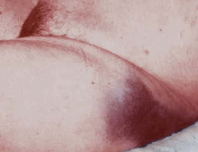
Non-traumatic ecchymosis over the upper outer aspect of the thigh secondary to abdominal haemorrhage. The ecchymosis is parallel with, but distal to the inguinal ligament with a well demarcated upper border defined by attachment of the membranous layer of the superficial fascia (Scarpa’s fascia).
Most often occurs in patients with retroperitoneal bleeding, usually due to acute haemorrhagic pancreatitis.
In 1966, John Adrian Fox detailed 2 fatal cases of non-traumatic ecchymosis determined as a diagnostic sign of retroperitoneal haemorrhage. In both cases, this sign was noticed late in the course and produced by tracking of the fluid extraperitoneally along the fascia of psoas and iliacus beneath the inguinal ligament until it became subcutaneous in the upper thigh
Case 1:
Fourteen hours after admission bruising was noted in both upper outer thighs. It had a sharp upper margin, was dark blue, and was quite distinct from the patchy mottling of her legs below
[Post mortem: acute suppurative pancreatitis]
Case 2:
A man, of about 50…with severe abdominal pain and circulatory collapse. Resuscitatory measures were of no avail and he died within 24 hours of admission. Before death bruising was noticed in the upper outer aspect of one thigh. He had not been given injections in this region and no other cause for the bruising was apparent.
[Post-mortem: dissecting and ruptured abdominal aortic aneurysm]
Proposed aetiology:
..seems likely that the clinical sign seen in the above 2 cases is produced by tracking of the fluid extraperitoneally along the fascia of psoas and iliacus beneath the inguinal ligament until it becomes subcutaneous in the upper thigh.
Cadaver Experiment:
This sign has been reproduced in two stages in the recent cadaver. A solution of methylene blue in normal saline was injected from a height of 10 feet into the loin for several hours. The blue dye was then traced by dissection until it was seen to pass beneath the inguinal ligament
Fox’s sign has been recorded in acute pancreatitis and ruptured aortic aneurysm and, by analogy with Cullen and Grey Turner signs, most probably occurs in any retroperitoneal loss of blood or bile.
Major Publications
- Fox JA. A diagnostic sign of extraperitoneal hemorrhage. Br J Surg. 1966; 53(3): 193-195
- Fox JA, Kreel L. Technique of retrograde colonic intubation and its initial application to high colonic biopsy. Gut. 1967; 8(1): 77-82
- Qvist G, Fox JA. Mucosal pyloroplasty. Br J Surg. 1969; 56(3): 172-177
- Fox JA. A fibreoptic colonoscope. Br Med J. 1969; 3(5661): 50
- Fox JA, Provenzale L, Revignas A. Fibreoptic colonoscopy. Lancet. 1970; 1(7663): 107.
References
- Epperla N, Mazza JJ, Yale SH. A Review of Clinical Signs Related to Ecchymosis. WMJ. 2015 Apr;114(2):61-5.
- Yale SH, Tekiner H, Yale ES. Recognition and confirmation of Fox sign. Forensic Sci Med Pathol. 2022 Mar;18(1):110-111.
- Cadogan M. Non-traumatic Abdominal Ecchymosis. LITFL 2020
Eponym
the person behind the name

Graduated MBBS St George's University of London in 2023 after completing my BSc in Human Genetics at University of Nottingham in 2018. I completed my foundation training at St. Mary's Hospital, Isle of Wight and achieved the national award for my contributions to the Enhance Enable Programme in Leadership. In 2025, I commenced my role in Emergency Medicine in Sir Charle's Gairdner Hospital, Perth, Western Australia.
BA MA (Oxon) MBChB (Edin) FACEM FFSEM. Emergency physician, Sir Charles Gairdner Hospital. Passion for rugby; medical history; medical education; and asynchronous learning #FOAMed evangelist. Co-founder and CTO of Life in the Fast lane | On Call: Principles and Protocol 4e| Eponyms | Books |