![]()
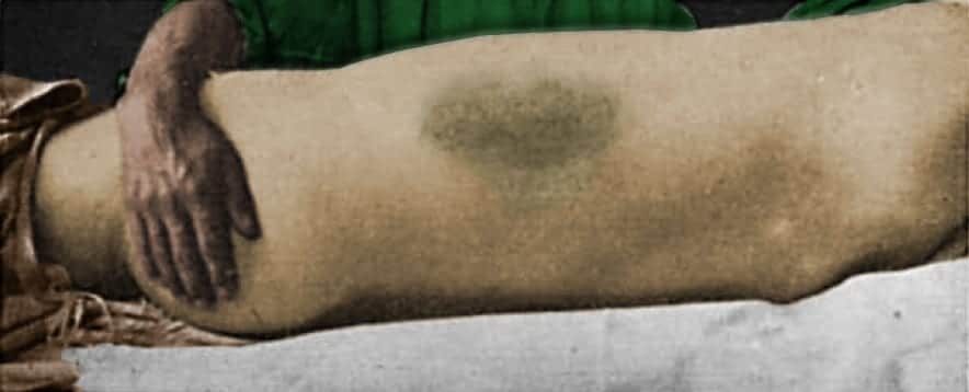
Grey Turner sign
Description
Grey Turner sign: atraumatic abdominal ecchymosis, in particular – bruising of the flanks. George Grey Turner reported a ‘dirty-green‘ discolouration on both loins of a patient with acute pancreatitis (confirmed on operation)
Incidence of 3-5% in patients with acute pancreatitis, associated with increased mortality (30-40%) and increased risk of pseudocyst formation. [Surg Gynecol Obstet. 1984]
CT scanning has helped to define the anatomic pathway by which extravasated pancreatic enzymes and their effects lead to these cutaneous discolouration. [Pancreas. 1998].
- Extraperitoneal diffusion is from the anterior pararenal space between the two leaves of the posterior renal fascia and then to the lateral edge of the quadratus lumborum muscle.
- Communication may then extend to the posterior pararenal space and the structures of the flank wall. The lumbar triangle, a site of anatomic weakness on the flank wall, provides an external window into the internal proteolytic events.
History of the Grey Turner sign
1920 – The English surgeon George Grey Turner (1877–1951) published paper on ‘Local discoloration of the abdominal wall as a sign of acute pancreatitis‘ citing two cases of acute pancreatitis with fat necrosis and retroperitoneal haemorrhage
The first case from 1912 concerned a 54 year old female with three days of abdominal pain presenting with an area of discoloration (a bluish colour), about 6 inches in diameter involving the abdominal wall surrounding the umbilicus.
…the patient suffered from acute pancreatitis, with much effusion into the peritoneal cavity. She lived nine days after operation, and the post-mortem examination disclosed a sloughing pancreas with much fat necrosis
Grey Turner 1920
The second case from 1917 prompted the observation “In a cursory examination of the voluminous literature on pancreatitis I have not observed any mention of this sign“
The tenderness over the gall-bladder region was very marked, and I now noticed two large discoloured areas in the loins. They were about the size of the palm of the hand, slightly raised above the surface, and of a dirty-greenish colour.
Grey Turner 1920
Alternate causes for Grey Turner Sign
| Cause | Reference |
|---|---|
| Intra-aortic balloon pump insertion | Rob, Williams 1961 |
| Cardiac catheterization | Armour et al 1978 |
| Sclerosing peritonitis | Pryor et al 2001 |
| Rectus sheath hematoma | Guthrie, Stanley 1996 |
| Ruptured abdominal aortic aneurysm | Armour et al 1978 |
| Retroperitoneal necrotizing fasciitis | Pryor et al 2001 |
| Ischemic and gangrenous bowel | Kelley ML 1961 |
| Bilateral acute salpingitis with IUP | Orient JM, Sapira JD 2005 |
| Acute Pancreatitis | Bosmann et al 2009 |
Epperla N, Mazza JJ, Yale SH. A Review of Clinical Signs Related to Ecchymosis. WMJ. 2015; 114(2): 61-5.
Associated Persons
- John Henry Bryant and Bryant sign (1903)
- Thomas Stephen Cullen and Cullen sign (1918)
- George Grey Turner and Grey Turner sign (1919)
- Francis Edward Stabler and Stabler Sign (1934)
- John Adrian Fox and Fox sign (1966)
Alternative names
- Grey Turner’s sign; Grey-Turner’s sign
Controversies
The topographic location of the ecchymosis of Grey Turner and Cullen do not point to the aetiology with any degree of certainty.
Grey Turner sign and Cullen sign may occur simultaneously, especially in patients with retroperitoneal haemorrhage.
References
Historical articles
- Grey Turner G. Local discoloration of the abdominal wall as a sign of acute pancreatitis. Br J Surg. 1920;7:394-395
Review articles
- Dickson AP, Imrie CW. The incidence and prognosis of body wall ecchymosis in acute pancreatitis. Surg Gynecol Obstet. 1984; 159(4): 343-7.
- Meyers MA, Feldberg MA, Oliphant M. Grey Turner’s sign and Cullen’s sign in acute pancreatitis. Gastrointest Radiol. 1989 Winter;14(1):31-7.
- Bem J, Bradley EL 3rd. Subcutaneous manifestations of severe acute pancreatitis. Pancreas. 1998; 16(4): 551-5.
- Mookadam F, Cikes M. Images in clinical medicine. Cullen’s and Turner’s signs. N Engl J Med. 2005; 353(13): 1386.
- Chung KM, Chuang SS. Cullen and Grey Turner signs in idiopathic perirenal hemorrhage. CMAJ. 2011 Nov 8;183(16):E1221
- Epperla N, Mazza JJ, Yale SH. A Review of Clinical Signs Related to Ecchymosis. WMJ. 2015; 114(2): 61-5.
- Chhabra P, Rana SS, Sharma V, Sharma R, Bhasin DK. Grey Turner’s sign in acute necrotizing pancreatitis. Ann Gastroenterol. 2015; 28(1): 147
FOAMed resources
- McKennedy C. Grey Turner sign: Eponym A Day. Instagram
- Cadogan M. Non-traumatic abdominal ecchymosis. 2020
[cite]
eponymictionary
the names behind the name
BA MA (Oxon) MBChB (Edin) FACEM FFSEM. Emergency physician, Sir Charles Gairdner Hospital. Passion for rugby; medical history; medical education; and asynchronous learning #FOAMed evangelist. Co-founder and CTO of Life in the Fast lane | On Call: Principles and Protocol 4e| Eponyms | Books |