Masquerading Bundle Branch Block (MBBB)
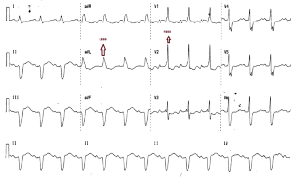
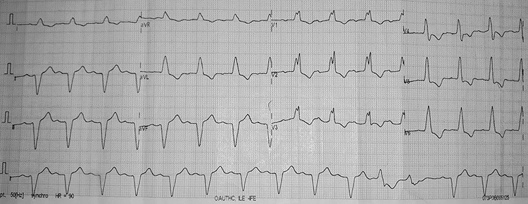
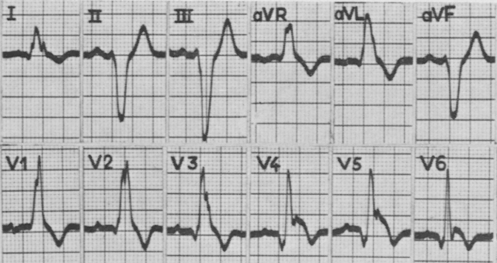
Mixed pattern of complete right bundle branch block (RBBB) in precordial leads and complete left bundle branch block (LBBB) in limb leads, indicating severe and diffuse conducting system disease with a poor prognosis
- Compared with typical bifascicular block, these patients have more extensive fibrosis and degeneration of left bundle pathways
- Consequent high-grade left anterior fascicular block (LAFB), often associated left ventricular enlargement, manifests as a complete LBBB. This is partially masqueraded by concurrent RBBB
- Patients have a higher risk of progression to complete AV block than typical bifascicular block
- A second, precordial type of MBBB has also been described (see below)
Diagnostic criteria
Standard or Precordial Masquerading Bundle Branch Block (MBBB). In both types, RBBB is shown by typical RSR’ pattern in lead V1.
Standard MBBB:
- RBBB pattern in precordial leads
- LBBB pattern in limb leads
- Small or absent S wave in lead I

Broad QRS complexes with an RBBB pattern (rsR pattern) in the precordial leads V1 to V3
LBBB pattern with near-absence of S waves in lead I in the limb leads.
Left axis deviation and QS complexes in Leads II, III, aVF and M pattern in lead aVL.
Precordial MBBB:
- RBBB pattern in leads V1-3
- LBBB pattern in V4-6
- Absent S wave in leads V5-6

RBBB pattern in right precordial leads with LBBB pattern in left precordial leads.
Absent S waves in V5-6 and lead I.
Clinical significance
Compared with a typical bifascicular block pattern, MBBB indicates more extensive disease of the left bundle pathway and left ventricle, and carries a poorer prognosis:
- Rate of progression to complete heart block over a four-year period in patients with MBBB was observed at 59% (Barrado et al), compared to 11% over a five-year period in typical bifascicular block (RBBB + LAFB)
- A large scale review of 600,000 ECGs found the rare MBBB pattern was associated with high mortality (41% over four years) and pacemaker insertion (39% over the same period)
- These observations were regardless of the presence or absence of trifascicular block
- Close follow-up of these patients and consideration of PPM insertion is essential even if asymptomatic
Differentiating from typical bifascicular block pattern relies mainly on the absence of prominent S waves in leads I and aVL.
Disease associations
- Ischaemic heart disease, in particular severe triple vessel disease
- Lenègre-Lev disease
- Chaga’s myocarditis
Associated persons
Justin L Richman and Louis Wolff first described this phenomenon in their 1954 publication in the American Heart Journal. They observed four cases of patients with ECGs with signs of LBBB in limb leads, and signs of RBBB in precordial leads. Subsequent vectorcardiograms revealed the nature of conduction deficit to be LBBB
Further examples
Example 1


Related Topics
References
- Richman JL, Wolff L. Left bundle branch block masquerading as right bundle branch block. Am Heart J. 1954; 47(3): 383-393.
- Lenègre J. Etiology and pathology of bilateral bundle branch block in relation to complete heart block. Prog Cardiovasc Dis 1964; 6: 409-444
- Gómez Barrado JJ, Turégano Albarrán S, García Rubira JC, Hidalgo Urbano R, Pavón García M, Berjillos Cordón M, Cruz Fernández JM. Características clínicas y electrocardiográficas del bloqueo bifascicular disfrazado [Clinical and electrocardiographic characteristics of masquerading bifascicular block]. Rev Esp Cardiol. 1997 Feb;50(2):92-7.
- Surawicz B, Knilans T. RBBB and Left Anterior Fascicular Block: Relation to Complete AV Block. In: Chou’s Electrocardiography in Clinical Practice: Adult and Pediatric 6e, 2008
- Maddali MM. Cardiac pacing in left bundle branch/bifascicular block patients. Ann Card Anaesth. 2010; 13(1): 7-15.
- Kaimoto et al. Masquerading bundle branch block as a marker of poor prognosis. J Cardiol Cases. 2013; 8(1): e57-59
- Elizari MV, Baranchuk A, Chiale PA. Masquerading bundle branch block: a variety of right bundle branch block with left anterior fascicular block. Expert Rev Cardiovasc Ther. 2013 Jan;11(1):69-75
- Schroder E Souza TG et al. Masquerading Bundle Branch Block: An Electrocardiographic Marker of Poor Prognosis. Circulation. 2015; 132(3): A14845.
- Dhanse et al. Masquerading Bundle Branch Block: A Poor Prognostic Sign Revisited. J Clin Diagn Res. 2016; 10(9): OD01–OD02.
- Jiao Z, Tian Y, Yang X, Liu X. Masquerading bundle branch block as a presenting manifestation of complete atrioventricular block that caused syncope. J Int Med Res. 2017; 45(5): 1597-1601
- Akinyele OA et al. Masquerading Bundle Branch Block in a Patient with Heart Failure in Nigeria. Int Med Case Rep J. 2020 Mar 30;13:105-110
Further reading
Online
- Wiesbauer F, Kühn P. ECG Mastery: Yellow Belt online course. Understand ECG basics. Medmastery
- Wiesbauer F, Kühn P. ECG Mastery: Blue Belt online course: Become an ECG expert. Medmastery
- Kühn P, Houghton A. ECG Mastery: Black Belt Workshop. Advanced ECG interpretation. Medmastery
- Rawshani A. Clinical ECG Interpretation ECG Waves
- Smith SW. Dr Smith’s ECG blog.
- Wiesbauer F. Little Black Book of ECG Secrets. Medmastery PDF
Textbooks
- Zimmerman FH. ECG Core Curriculum. 2023
- Mattu A, Berberian J, Brady WJ. Emergency ECGs: Case-Based Review and Interpretations, 2022
- Straus DG, Schocken DD. Marriott’s Practical Electrocardiography 13e, 2021
- Brady WJ, Lipinski MJ et al. Electrocardiogram in Clinical Medicine. 1e, 2020
- Mattu A, Tabas JA, Brady WJ. Electrocardiography in Emergency, Acute, and Critical Care. 2e, 2019
- Hampton J, Adlam D. The ECG Made Practical 7e, 2019
- Kühn P, Lang C, Wiesbauer F. ECG Mastery: The Simplest Way to Learn the ECG. 2015
- Grauer K. ECG Pocket Brain (Expanded) 6e, 2014
- Surawicz B, Knilans T. Chou’s Electrocardiography in Clinical Practice: Adult and Pediatric 6e, 2008
- Chan TC. ECG in Emergency Medicine and Acute Care 1e, 2004
ECG LIBRARY
MBBS DDU (Emergency) CCPU. Adult/Paediatric Emergency Medicine Advanced Trainee in Melbourne, Australia. Special interests in diagnostic and procedural ultrasound, medical education, and ECG interpretation. Co-creator of the LITFL ECG Library. Twitter: @rob_buttner


