
![]()
Max Brödel
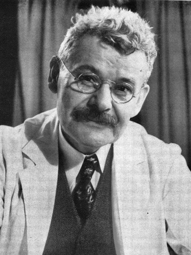
Max Brödel (1870–1941) was a German medical illustrator, educator, and pioneer of medical art
Brödel’s mastery of illustration transformed medical education and practice. Regarded as the founder of modern medical illustration, he combined artistic talent with a deep understanding of anatomy and surgery to create visuals that conveyed far more than any photograph could achieve. His philosophy—that medical artists must first acquire comprehensive knowledge of anatomy, pathology, and surgical technique—laid the foundation for a new profession.
Arriving in Baltimore in 1894, Brödel joined the Johns Hopkins Hospital at the invitation of Franklin P. Mall and quickly became an indispensable collaborator to leading surgeons such as Howard A. Kelly, William S. Halsted, Thomas S. Cullen, and Harvey Cushing. His illustrations for Kelly’s Operative Gynecology (1898) set a new standard for medical texts and secured Brödel’s reputation worldwide. Beyond his technical skill, he was an innovator, introducing techniques such as the carbon dust method to achieve lifelike depictions of tissue.
Brödel’s career reached its zenith in 1911 when he founded the Department of Art as Applied to Medicine at Johns Hopkins, the first program of its kind in the world. As its first director and associate professor, he trained a generation of medical illustrators, including James Didusch and his own daughter, Elizabeth Brödel. His graduates went on to disseminate his principles globally, influencing medical education for decades.
A man of artistic temperament and musical passion, Brödel was also a keen investigator. When surgical details were unclear, he performed dissections and experiments to ensure accuracy in his drawings—work that led to anatomical insights such as the avascular plane of the kidney and the development of Brödel’s suture for nephropexy. His illustrations were not mere images but acts of scientific interpretation, guiding clinicians and students alike.
Biographical Timeline
- Born June 8, 1870 in Leipzig, Germany, to Louis Brödel and Henrietta Frenzel.
- 1884–1885 Technical High School, Leipzig.
- 1885–1890 Studied at Leipzig Academy of Fine Arts; began work in anatomical and physiological institutes under Wilhelm His, Braune, and Carl Ludwig.
- 1890 – Drafted into German Army; served one year with arms, second year doing artistic work for his regiment through influence of Carl Ludwig and Prince George of Saxony.
- 1892 – Returned to Leipzig as freelance artist specializing in anatomical illustrations.
- 1894 – Recruited by Franklin P. Mall (1862–1917), anatomist and embryologist to Johns Hopkins. Arrived in Baltimore (Jan 18) and began collaboration with Howard A. Kelly.
- 1895 – Joined by Hermann Becker and later August Horn as illustrators in Kelly’s department
- 1898 – Published illustrations in Kelly’s Operative Gynecology (2 vols), established Brödel’s reputation and revolutionized medical illustration.
- 1900 – Colour reproductions of Brödel’s work in Cancer of the Uterus marked a high point in medical illustration quality.
- 1902 – Married fellow illustrator Ruth Huntington (Dec 31); later fathered four children (Elizabeth, Ruth, Carl, Elsa).
- 1909 – Elected Honorary Member of Medical and Chirurgical Faculty of Maryland—the only layperson to receive this honour.
- 1910 – Invited to join staff at Mayo Clinic; Thomas Cullen persuaded Henry Walters to fund creation of the Department of Art as Applied to Medicine at Johns Hopkins to keep Brödel in Baltimore.
- 1911 – Appointed Associate Professor and first Director of the Department of Art as Applied to Medicine at Johns Hopkins, the first program of its kind in the world.
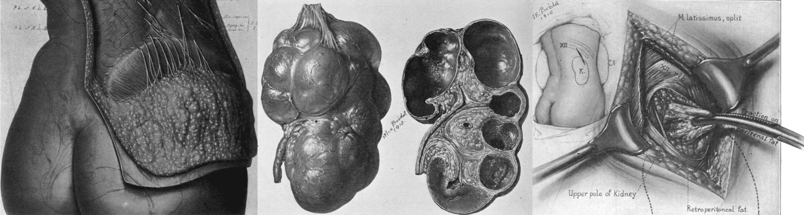
- 1914 – Contributed 357 illustrations to Kelly & Burnham’s Diseases of the Kidneys, Ureters and Bladder a landmark publication in urology
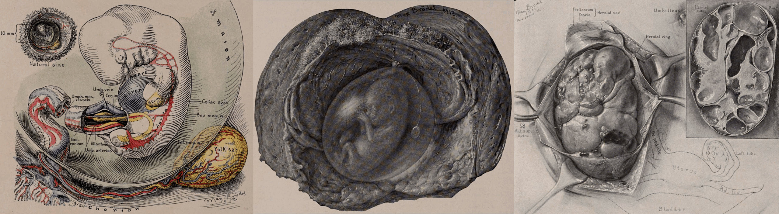
- 1916 – Produced embryology illustrations for Cullen’s Embryology, Anatomy, and Diseases of the Umbilicus; utilized the Carnegie Collection of Human Embryos for detailed studies.
- 1920s–1930s – Continued leadership of the Hopkins department; trained many prominent medical illustrators including James F. Didusch (1890-1955) and daughter Elizabeth H. Brödel (1903-1986).
- 1933 – Delivered address at Howard Kelly’s 75th birthday celebration, reflecting on their pioneering work together.
- Published landmark article “Medical illustration” in JAMA (Aug 30); died October 26, 1941 in Baltimore of pancreatic cancer, aged 71.
Medical Eponyms
Brödel’s Suture
Brödel’s suture is a triangular, mattress-style fixation suture used in nephropexy (surgical fixation of a mobile or prolapsed kidney). It provides secure anchoring by distributing tension across a wider surface of renal tissue, reducing the risk of tearing through the parenchyma.
The technique was developed during Brödel’s early years at Johns Hopkins (1894–1902) while illustrating for Howard A. Kelly. The idea originated from his anatomical injection and dissection studies on kidney vascular supply, which revealed an avascular zone suitable for incision and fixation.
First described in a short communication in American Medicine (1902), where Brödel published A more rational method of passing the suture in fixation of the kidney.
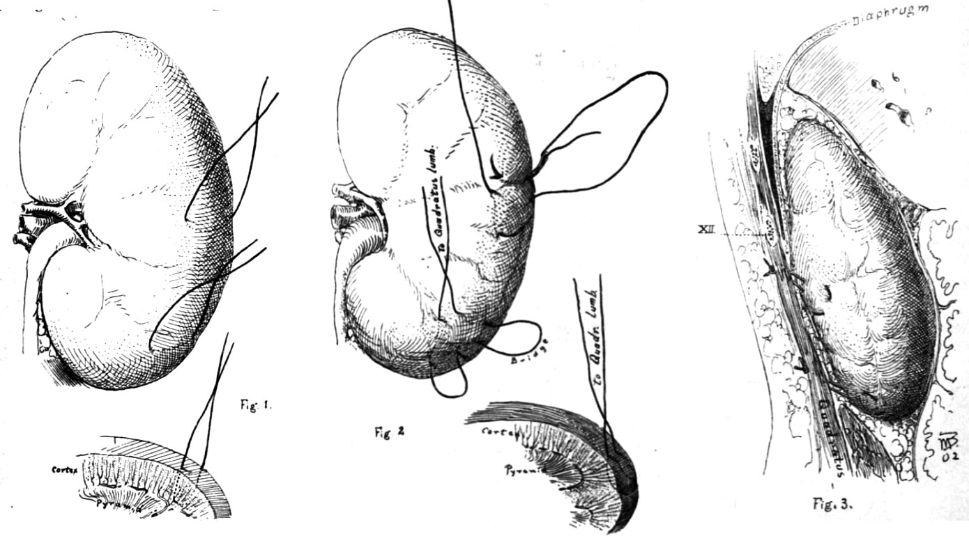
Although nephropexy techniques have evolved, Brödel’s anatomical insight and methodical approach remain significant in urologic surgery. His name persists in surgical history for linking artistic observation with operative innovation.
Illustration Techniques & Innovations
- Introduced carbon dust technique for half-tone realism and depth.
- Combined multiple views, magnifications, and operative steps in single didactic compositions.
- Advocated exhaustive pre-illustration research
It should be remembered that technique, artistic feeling, accurate draftsmanship, neatness and speed are all relatively unimportant. The planning of the picture and the registration of the scientific facts are what gives it its value, not the execution…To make such a picture the artist must know his subject so thoroughly that he can shut his eyes and coax into existence a mental picture of great clarity, complete in every respect.
Notable Collaborations
Howard A. Kelly (1858–1943)
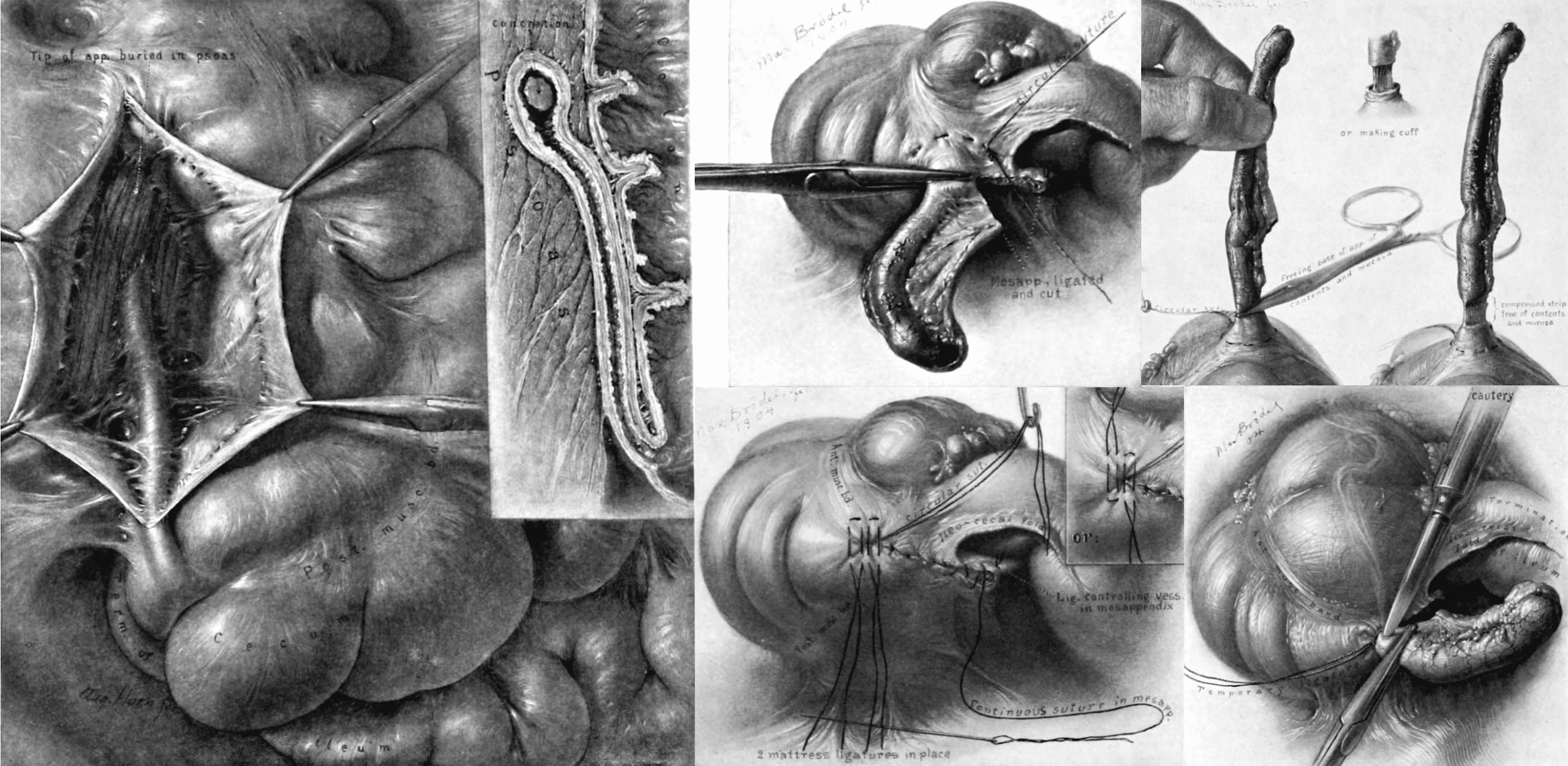
Chief of Gynecology at Johns Hopkins; Brödel illustrated Kelly’s Operative Gynecology (1898-1903), providing over 367 images that revolutionized gynecological education. Later worked on The Vermiform Appendix and Its Diseases (1905; 117 illustrations); Diseases of the Kidneys, Ureters and Bladder (1914; 450 illustrations); and Medical Gynecology (1911).
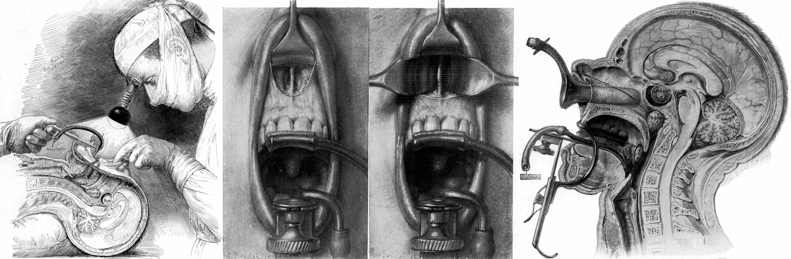
Harvey Cushing (1869–1939)
Collaborated on early neurosurgical illustrations; e.g., Surgical Experiences with Pituitary Disorders (1914) which featured Brödel’s ink drawings for neurosurgical approaches. Cushing trained in Brödel’s studio and their correspondence reflects their close working relationship.
Thomas S. Cullen (1868–1953)
Provided colour illustrations to greatly enhance the texts of Cancer of the Uterus (1900); Embryology, Anatomy, and Diseases of the Umbilicus (1916) and monographs on myomata and rectus muscle lesions.
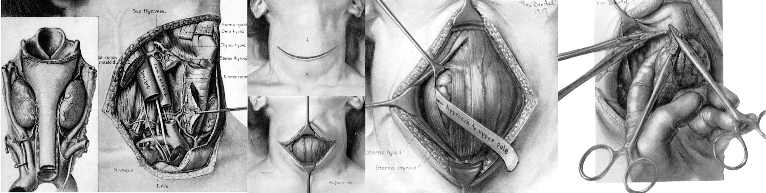
William S. Halsted (1852–1922)
Johns Hopkins surgeon; Brödel’s injection studies enabled Halsted to describe parathyroid vascular supply (1917) and informed thyroid surgery.
Major Publications
- Brodel M. The intrinsic blood-vessels of the kidney and their significance in nephrotomy. Bulletin of the Johns Hopkins Hospital 1901; 12(118): 10-13
- Brodel M. A more rational method of passing the suture in fixation of the kidney. American Medicine 1902; 4: 176-178
- Brodel M. How may our present methods of medical illustration be improved. JAMA 1907;XLIX;(2):138-140.
- Brodel M. Testimonial dinner to Howard Atwood Kelly on his seventy-fifth birthday. Bull Johns Hopkins Hosp 1933; 53(2): 65-109.
- Brodel M. Medical illustration. JAMA 1941; 117(9): 668-72.
- Kelly HA, Burnham CF. Diseases of the kidneys, ureters and bladder, New York: D. Appleton, 1914. [Volume 2, 1922]
References
Biography
- Cullen TS. Max Brödel, 1870-1941 Director of the First Department of Art as Applied to Medicine in the World. Bull Med Libr Assoc. 1945 Jan;33(1):4.1–29.
- Engel RM. Max Brodel (1870-1941). Invest Urol 1969;7(2):192-3.
- Crosby RW, Cody J. Max Brödel. The Man Who Put Art Into Medicine. 1991
- Wolff M, Radwan H. Max Brödel (1870-1941): sein Leben und Beitrag zur Entwicklung der modernen Chirurgie [Max Brödel (1870-1941): his life and contribution to the development of modern surgery]. Chirurg. 1997 Aug;68(8):840-7.
- Schultheiss D, Jonas U. Max Brödel (1870-1941) and Howard A. Kelly (1858-1943)–urogynecology and the birth of modern medical illustration. Eur J Obstet Gynecol Reprod Biol. 1999 Sep;86(1):113-5.
- Schultheiss D, Engel RM, Crosby RW, Lees GP, Truss MC, Jonas U. Max Brödel (1870-1941) and medical illustration in urology. J Urol. 2000 Oct;164(4):1137-42.
- Max Brödel (1870-1941). Embryology History
- Max Brödel (1870-1941). William P. Didusch Center for Urologic History
Eponym
the person behind the name

BA MA (Oxon) MBChB (Edin) FACEM FFSEM. Emergency physician, Sir Charles Gairdner Hospital. Passion for rugby; medical history; medical education; and asynchronous learning #FOAMed evangelist. Co-founder and CTO of Life in the Fast lane | On Call: Principles and Protocol 4e| Eponyms | Books |