![]()
Osler node
Osler nodes are painful, erythematous or violaceous, raised nodules typically found on the pads of the fingers and toes, often with a central pallor. They are one of the classic peripheral stigmata of infective endocarditis (IE), most often associated with subacute presentations and likely caused by immune complex deposition or septic emboli. Their onset is often preceded by localized pain, and the lesions typically resolve within hours to days without scarring.
Although historically considered distinct from Janeway lesions — which are non-tender and embolic — modern studies reveal histopathologic overlap. Osler nodes may contain neutrophilic microabscesses and occasionally yield bacterial cultures, suggesting a spectrum of septic microvascular skin involvement in endocarditis.
Named after Sir William Osler (1849–1919), the lesions were highlighted in his landmark 1885 Gulstonian Lectures on malignant endocarditis. However, earlier accounts by John A. Mullen and French clinicians describe similar lesions. Osler’s prominence and diagnostic emphasis likely led to the widespread adoption of the eponym.
Description
Osler node: Painful, red, raised lesions usually found on the palms and soles. Caused by immune complex deposition and the resulting inflammatory response.
Associated with a number of conditions, including infective endocarditis, SLE, and disseminated gonococcal infection. To be differentiated from painless Janeway lesions
History of Osler nodes
1873 – Armand Trousseau and Germain Sée report painful nodules in patients with ulcerative endocarditis, later termed nodosités cutanées éphémères.
Late 1800s – John Alexander Mullen (1835–1899), a Canadian physician, documents tender subcutaneous nodules in cases of endocarditis in Hamilton, Ontario.
1885 – Sir William Osler (1849-1919), in a series of three Gulstonian lectures on malignant endocarditis described the cutaneous manifestations of severe infectious endocarditis as
haemorrhages…upon the skin, serous and mucous surfaces…in which a minute necrotic or suppurative centre can sometimes be seen.
Osler 1885
1888 – Osler’s attention drawn to the ephemeral spots of a painful nodular erythema by John Alexander Mullin (1835–1899) of Hamilton, Ontario whose description he found admirable and later quoted in his 1909 publication
The spots came out at intervals as small swollen areas, some the size of a pea, others a centimetre and a half in diameter, raised, red, with a whitish point in the centre. I have known them to pass away in a few hours, but more commonly they last for a day, or even longer. The commonest situation is near the tip of the finger, which may be slightly swollen.
Mullen JA, 1888
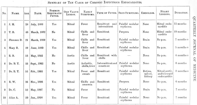
1908 – Osler presented to the Association of Physicians of Great Britain and Ireland in July 1908 (published 1909). He reported on ten patients with chronic infectious endocarditis, including seven with cutaneous lesions.
One of the most interesting features of the disease (chronic endocarditis) and one to which very little attention has been paid is the occurrence of ephemeral spots of a painful nodular erythema, chiefly in the skin of the hands and feet, des nodosités cutanées éphémères of the French. The blood-cultures and the presence of the painful erythematous nodules and the occurrence of embolism furnish the most important aids.
Osler went on to explain how nodules of this character occurred in seven of the cases, and in three at least they were of importance in determining the diagnosis.
…the presence of these spots appeared to me to clinch the diagnosis. They are not beneath but in the skin and they are not unlike an ordinary wheal of urticaria. The pad of the finger and toe , the thenar and hyperthenar eminences, the sides of the fingers, and the skin of the over part of the arm are the most common localities. In one case they were present in the skin of the flank. I have never seen them haemorrhagic, but always erythematous, sometimes of a very vivid pink hue, with a slightly opaque centre.
1990 – Cardullo et al. show histologic similarity between Osler nodes and Janeway lesions — both may reveal neutrophilic dermal abscesses, sometimes culture-positive for bacteria.
2008 – Marrie reviews Osler nodes as a useful but rare diagnostic clue for IE, present in fewer than 10% of modern cases.
2023 – Buchanan et al. clarify historical attribution, asserting Mullen first described these lesions but Osler gave them clinical diagnostic prominence.
Associated Persons
- Armand Trousseau, MD (1801–1867) – Early reference to painful cutaneous nodules in systemic illness; contributed to broader knowledge of embolic signs
- Edward Gamaliel Janeway (1841-1911) [Janeway lesion]
- Emanuel Libman (1872-1946)
- John Alexander Mullin (1835–1899)
- Sir William Osler (1849 -919) [Osler nodes]
References
Original articles
- Osler W: Gulstonian lectures on malignant endocarditis. Lancet. 1885; 125(3210): (I 415-418), (II 459-464), (III 505-508)
- Osler W. Chronic infectious endocarditis. Quarterly Journal of Medicine. 1909; 2(2): 219–230.
Review articles
- Farrior JB, Silverman ME. A consideration of the differences between a Janeway’s lesion and an Osler’s node in infectious endocarditis. Chest. 1976 Aug;70(2):239-43.
- Cardullo AC, Silvers DN, Grossman ME. Janeway lesions and Osler’s nodes: a review of histopathologic findings. J Am Acad Dermatol. 1990 Jun;22(6 Pt 1):1088-90.
- Marrie TJ. Osler’s nodes and Janeway lesions. Am J Med. 2008 Feb;121(2):105-6
- Sethi K, Buckley J, de Wolff J. Splinter haemorrhages, Osler’s nodes, Janeway lesions and Roth spots: the peripheral stigmata of endocarditis. Br J Hosp Med (Lond). 2013 Sep;74(9):C139-42.
- Servy A et al; Association Pour l’Etude et la Prévention de l’Endocardite Infectieuse Study Group. Prognostic value of skin manifestations of infective endocarditis. JAMA Dermatol. 2014 May;150(5):494-500.
- Gomes RT et al. Dermatologic manifestations of infective endocarditis. An Bras Dermatol. 2016 Sep-Oct;91(5 suppl 1):92-94.
- Buchanan WW, Rainsford K, Kean CA, Kean WF. John Alexander Mullin (1835–1899): the Canadian physician who first described Osler’s nodes. Inflammopharmacology. 2023
- Parashar K, Daveluy S. Osler Node and Janeway Lesions. 2023 Jul 24. In: StatPearls
eponymictionary
the names behind the name
Doctor in Australia. Keen interest in internal medicine, medical education, and medical history.
BA MA (Oxon) MBChB (Edin) FACEM FFSEM. Emergency physician, Sir Charles Gairdner Hospital. Passion for rugby; medical history; medical education; and asynchronous learning #FOAMed evangelist. Co-founder and CTO of Life in the Fast lane | On Call: Principles and Protocol 4e| Eponyms | Books |