![]()
Paul Tillaux
Paul Jules Tillaux (1834-1904) was a French Surgeon.
Tillaux performed cadaveric experiments and demonstrated stress applied to the anterior inferior tibiofibular ligament resulted in an avulsion fracture.
Eponymously affiliated with multiple eponyms most notably the Aïe crépitant de Tillaux (1895); Manœuvre de Tillaux; Appareil de Tillaux; the Tillaux fracture; the Spiral of Tillaux; and the Tillaux enucleation “from behind forward“
A similar injury to the anterolateral tibia cadeveric description by Tillaux (1872) was later described by Henri Chaput in 1907 hence the term the Tillaux-Chaput fracture.
Biography
- Born on December, 8 1834 in Aulnay-sur-Odon, Calvados
- 1857 – Interne des Hôpitaux
- 1862 – Doctor of medicine, Paris. Theses: ‘Des conduits excréteurs des glandes sublinguale et lacrymale‘ [Excretory ducts of the sublingual and lacrimal glands
- 1863 – Surgeon to the Bureau Central
- 1866 – Professeur-Agrégé of Surgery, in Paris and defended his aggregation entitled: Des Affections chirurgicales des Nerfs
- 1868 – Director of the Amphitheatre d’Anatomie des Hopitaux de Paris
- 1878 – Surgeon to the Beaujon Hospital
- 1890 – Professor of Clinical Surgery at the Hôpital de la Charité
- 1900 – Hon FRCS at the Royal College of Surgeons, England
- 1904 – President of the Académie de Médecine
- Died on October 20, 1904
Medical Eponyms
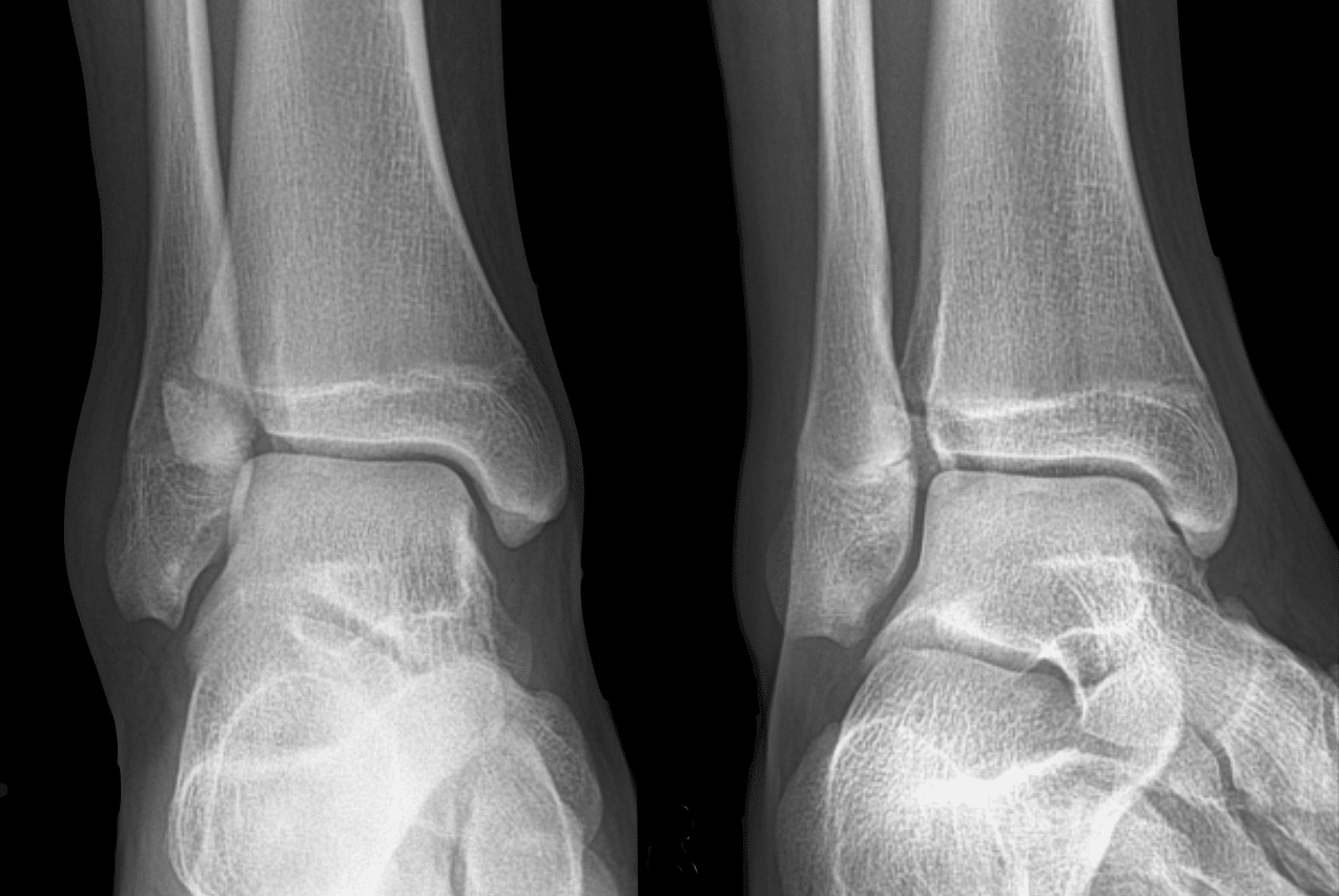
Tillaux fracture (1872) [Tillaux-Chaput fracture]
Fracture of the anterolateral tibial epiphysis commonly seen in adolescents. (Salter-Harris III tibial fracture)
Usually associated with forced lateral rotation of the foot or medial rotation of the leg on a fixed foot. This rotational injury results in avulsion of the anterior tibiofibular ligament at the lateral epiphysis. Often misdiagnosed as a simple sprain in adolescents.
Best seen on conventional AP, lateral, and mortise ankle XR. However, CT scan has better sensitivity in diagnosing Tillaux fractures and used to detect fracture displacement of >2mm – often an indication for open reduction
Orthopaedic eponyms
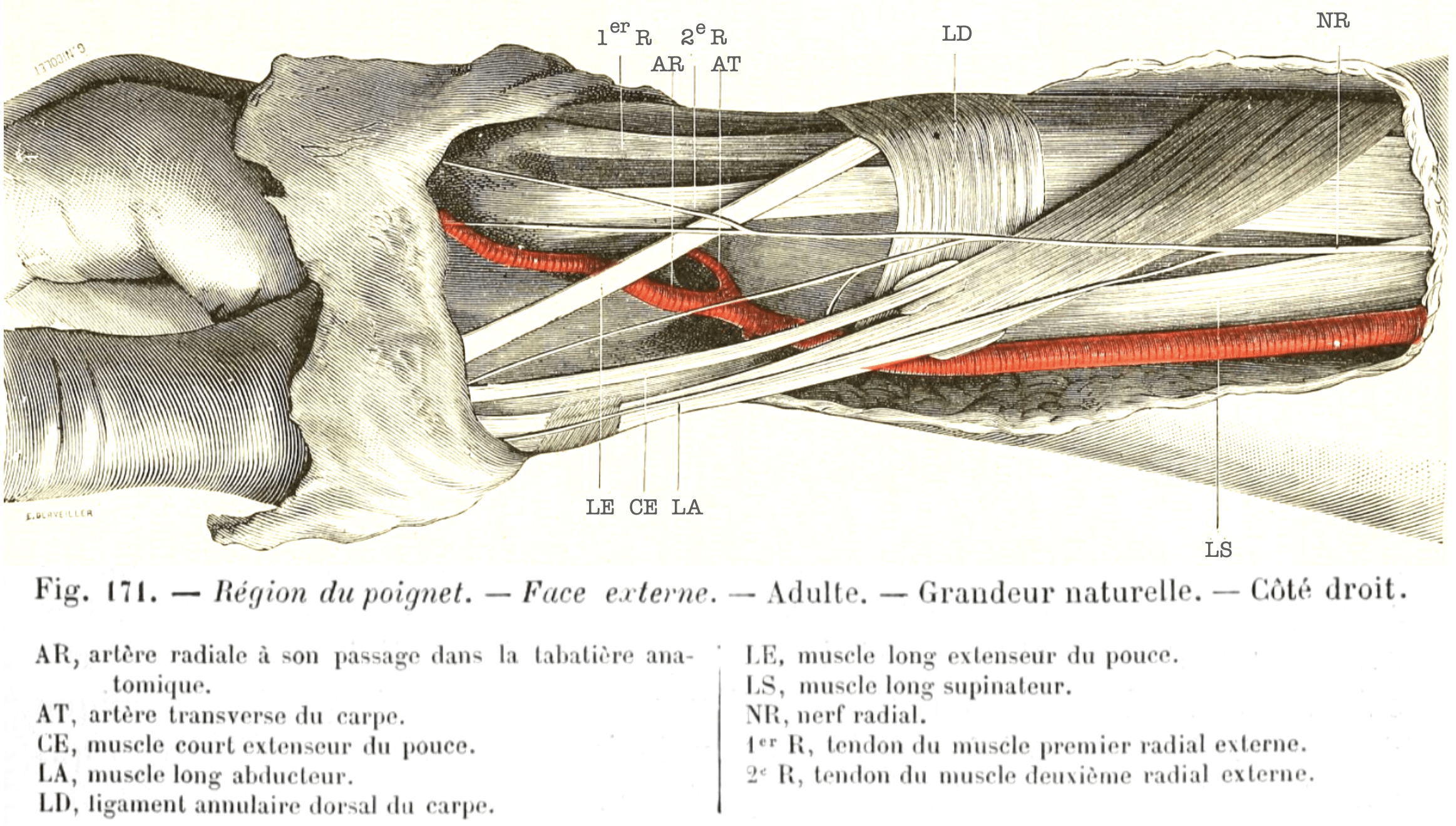
Tillaux painful crepitus sign (Aïe crépitant de Tillaux)
Tillaux described ténosite crépitante ou d’aï of the tendon sheaths of the thumb, particularly the long abductor and short extensor. This painful crepitating tenosynovitis later became associated with the term Aïe crépitant de Tillaux before de Quervain description in 1895
Original
English
Les coulisses destinées à loger les tendons du pouce, et en particulier celles du long abducteur et du court extenseur, sont fréquemment le siège d’une inflammation désignée sous le nom de ténosite crépitante ou d’aï…Elle se traduit par un gonflement siégeant sur le trajet de ces tendons et un bruit spécial lorsque le malade remue le pouce…Tillaux 1877
The sheaths intended to lodge the tendons of the thumb, and in particular those of the long abductor and short extensor, are frequently the seat of an inflammation designated by the name crepitating tenositis or ‘aï’…It is manifested by swelling along the course of these tendons and a special noise when the patient moves the thumb…Tillaux 1877
Tillaux apparatus (Appareil de Tillaux)
Apparatus used to reduce and maintain femoral diaphysis fractures in constant steady extension. The device was made up of strips of diachylon, glued to the outer and inner sides of the leg, forming a stirrup under the foot. To this stirrup, the traction cord was attached.

Other eponyms
Tillaux manoeuvre (Manœuvre de Tillaux)
Clinical examination of the breast: to demonstrate the adhesion of a breast tumor to pectoralis major. The mobility of the tumor within the deep planes is reduced when pectoralis major is contracted, by opposing adduction of patients arm.
Spiral of Tillaux
An imaginary line connecting the insertions of the recti muscles of the eye in relation to the limbus.
Conventional teaching states that the spiral of Tillaux marks the location of the ora serrata. On literature review, no source for this was found.

Tillaux enucleation from behind forward
Tillaux introduced his own modified technique for the enucleation of the eye
Original
English
Diviser la conjonctive et le fascia sous-conjonctival avec des ciseaux courbes, au niveau de l’attache à la sclérotique du muscle droit externe ; diviser le tendon de ce muscle et porter immédiatement les ciseaux par la boutonnière conjonctivale jusque sur le nerf optique; diviser ce nerf à son entrée dans le globe oculaire; saisir le pôle postérieur du globe avec une pince à griffes et l’attirer en dehors à travers la boutonnière conjonctivale, achever ensuite l’opération, en rasant la sclérotique.
Ce mode d’extraction s’opère avec une grande rapidité. Plus que les autres procédés, il diminue les chances d’ouverture de la loge postérieure de l’orbite.
Divide the conjunctiva and subconjunctival fascia with curved scissors, at the level of the attachment to the sclera of the external rectus muscle; divide the tendon of this muscle and immediately bring the scissors through the conjunctival buttonhole to the optic nerve; divide this nerve as it enters the eyeball; grasp the posterior pole of the globe with clawed forceps and draw it out through the conjunctival buttonhole, then complete the operation, shaving off the sclera.
This mode of extraction takes place with great rapidity. More than the other processes, it decreases the chances of opening the posterior compartment of the orbit.
Major Publications
- Tillaux PJ. Des conduits excréteurs des glandes sublinguale et lacrymale: du rôle des sinus de la face. Thèse pour le doctorat en médecine, 4 février 1862
- Tillaux PJ. De l’urétrotomie. [Urethrotomy] Asselin 1863
- Tillaux PJ. Des Affections chirurgicales des Nerfs, 4th, Paris, 1866
- Tillaux PJ. Recherches cliniques et expérimentales sur les fractures malléolaires. [Reported by Leon Gosselin]. Bulletin de l’Academie de médecine. 1872; 21: 817-826 [Tillaux fracture]
- Tillaux PJ. Recherches expérimentales et cliniques sur le mécanisme de la production des luxations coxo-fémorales en arrière. 1876
- Tillaux PJ. Traité d’anatomie topographique avec applications à la chirurgie. 1877 [Dedicated to Prof Gosselin] [2e 1879, 3e 1882, 4e 1884 5e 1887, 6e 1890]
- Tillaux PJ. Trattato di anatomia topografica : con applicazioni alla chirurgia. 1885
- Tillaux PJ. Traité de chirurgie clinique. 1888, [Volume II]
References
Biography
- Obituary: Paul Tillaux, M.D. Br Med J 1904; 2: 1196
- Rémy F . Un chirurgien du 19e siécle: Paul Tillaux. Thése d’exercice: Médecine. Université de Caen. 1985.
- Konofaou V, Dafereras M, Georgakopoulos P, Mavrommatis E. Professor Paul Jules Tillaux (1834–1904): His Contribution to Surgery and His Unknown Pioneer Work in Ophthalmic Surgery. Surgical Innovation. April 2021.
- Biography: Tillaux, Paul Jules (1834 – 1904). Plarr’s Lives of the Fellows Online. Royal College of Surgeons of England.
- Bibliography. Tillaux, Paul Jules 1834-1904. WorldCat Identities
Eponymous terms (orthopaedic)
- Lebrun A. L’appareil de Tillaux dans les fractures de la diaphyse du fémur. Bruxelles: 1880
- Protas JM, Kornblatt BA. Fractures of the lateral margin of the distal tibia. The Tillaux fracture. Radiology. 1981 Jan;138(1):55-7.
- Dias LS, Giegerich CR. Fractures of the distal tibial epiphysis in adolescence. J Bone Joint Surg Am. 1983 Apr;65(4):438-44
- Koury SI, Stone CK, Harrell G, La Charité DD. Recognition and management of Tillaux fractures in adolescents. Pediatr Emerg Care. 1999 Feb;15(1):37-9
- Konofaou V, Dafereras M, Georgakopoulos P, Mavrommatis E. Professor Paul Jules Tillaux (1834-1904): His Contribution to Surgery and His Unknown Pioneer Work in Ophthalmic Surgery. Surg Innov. 2021 Apr
- Botz B. Tillaux fracture Case. Radiopaedia
- Hacking C. Tillaux fracture. Radiopaedia
- McKennedy C. Tillaux fracture – Eponym A Day. Instagram
- Eponymythology: Eponymous Foot, ankle and talus injuries. LITFL
Eponymous terms (ophthalmology)
- Lefert P. La pratique des maladies des yeux dans les hôpitaux de Paris: aide-mémoire et formulaire de thérapeutique appliquée. 1895: 136-139
- Beard CH. Ophthalmic Surgery; a Treatise on Surgical Operations Pertaining to the Eye and its Appendages. 1914: 483
- White MH, Lambert HM, Kincaid MC, Dieckert JP, Lowd DK. The ora serrata and the spiral of Tillaux. Anatomic relationship and clinical correlation. Ophthalmology. 1989 Apr;96(4):508-11.
Eponym
the person behind the name
Dr Josh Howard - wannabe future orthopod, finding myself in Australia | LinkedIn
BA MA (Oxon) MBChB (Edin) FACEM FFSEM. Emergency physician, Sir Charles Gairdner Hospital. Passion for rugby; medical history; medical education; and asynchronous learning #FOAMed evangelist. Co-founder and CTO of Life in the Fast lane | On Call: Principles and Protocol 4e| Eponyms | Books |