


Westermark sign
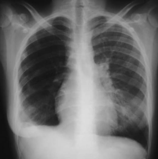
Westermark sign describes regional pulmonary oligaemia on chest radiography distal to pulmonary arterial obstruction by an embolus. The affected region appears relatively hyperlucent, with attenuation or absence of peripheral pulmonary vascular markings and sometimes abrupt termination of the normally visible central vessels as they enter the hypoperfused lung.
The appearance primarily reflects reduced pulmonary arterial perfusion distal to the embolic obstruction, potentially accentuated by peripheral pulmonary arterial vasoconstriction. The extent may range from a segmental or wedge-shaped region to an entire lobe or lung, depending on the level and extent of vascular obstruction.
Westermark sign is uncommon and has low sensitivity for acute pulmonary embolism. When present in an appropriate clinical setting it is a useful radiographic clue, but its absence does not exclude PE and definitive diagnosis requires appropriate vascular imaging.
Westermark sign may raise suspicion for PE when present, but its absence has little exclusionary value.
Historical development
1938 – Nils Westermark published On the roentgen diagnosis of lung embolism. Since 1933 he had examined 26 patients radiographically in whom pulmonary arterial embolism was demonstrated at autopsy. All had been radiographed within two weeks of death, many repeatedly. Four were imaged on the day of death and eight within two days.
Of these 26 patients, 10 showed pulmonary infarction, while the remaining 16 demonstrated embolism without infarction. Importantly, in seven of the ten infarction cases the radiographic evidence of embolic vascular obstruction involved a larger territory than the infarct itself. Westermark then studied a further 18 clinical cases, comparing radiographs during the embolic episode with films obtained during healing and recovery.
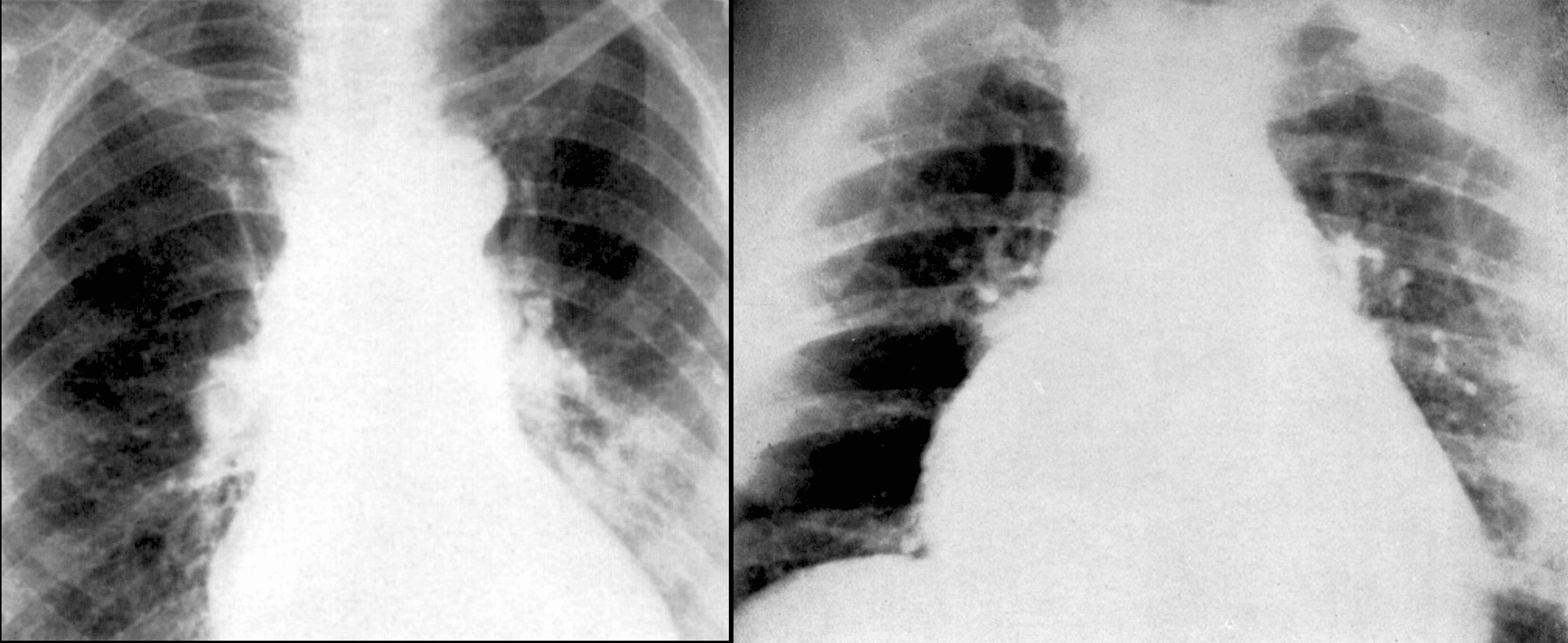
In embolism without infarction, the pulmonary arterial branches distal to the embolus were depleted of blood and contracted. The corresponding lung remained aerated, producing a localised area of increased lucency with attenuated or absent vascular markings. The central vessels remained visible but could terminate abruptly as they reached the oligaemic territory.
In embolism of the pulmonary artery without infarction we get ischaemia of the branches of the pulmonary artery on the peripheral side of the embolus. On the radiogram this ischaemia appears as a clarified area with diminished vascular design corresponding to the extent of the embolised artery. The vascularisation is however maintained in the central parts of the lung. The vascularisation takes however a rapid end to pass over into the above mentioned area of non-vascularisation.
Westermark 1938
1951 – Hanelin and Eyler published Pulmonary Artery Thrombosis: Roentgen Manifestations reviewing five cases involving the main pulmonary artery or major branches. They credited Westermark with describing decreased peripheral vessel visibility and increased radiolucency, together with enlargement and apparent abrupt termination of major vascular shadows.
1977 – Bedard and Bone published Westermark’s sign in the diagnosis of pulmonary emboli in patients with the adult respiratory distress syndrome. They described two patients with ARDS in whom development of localised hyperlucency against otherwise diffuse pulmonary infiltrates prompted recognition of superimposed PE, subsequently confirmed by pulmonary arteriography.
1993 – Worsley et al., analysing the PIOPED cohort, prospectively evaluated the classic CXR findings of acute PE. Westermark sign proved relatively specific but poorly sensitive, with reported values of approximately 14% sensitivity and 92% specificity.
2007 – Sreenivasan, Bennett and Parfitt published a modern correlation. CXR demonstrated focal right-sided oligaemia together with Palla sign, while perfusion scintigraphy showed a corresponding large perfusion defect in the same region.
2014 – Abbas et al. demonstrated Westermark and Palla signs on CXR with CTPA showing subtotal right main pulmonary arterial occlusion and marked relative oligaemia of the right upper and middle lobes.
2020 – Al Dandan et al. published Clinical and imaging profiles of pulmonary embolism. Among 83 patients with PE who had a chest radiograph, only 2 (2.4%) demonstrated Westermark sign. The study contains only confirmed PE cases and cannot estimate specificity, but reinforces how rarely the sign is encountered in contemporary practice.
Eponymythology
Westermark did not describe a “Westermark sign”. His 1938 paper described a clarified area with diminished vascular design caused by pulmonary arterial embolism without infarction. The eponymous term was applied subsequently.
Modern descriptions often define Westermark sign simply as unilateral or regional hyperlucency. Westermark’s original description was more specific “reduced or absent peripheral vascular markings corresponding to an embolised arterial territory, with preservation and abrupt termination of central vessels“. Hyperlucency was therefore the consequence of regional vascular depletion, not the defining observation in isolation.
Associated Persons
- Nils Westermark (1892-1980) [Westermark sign]
- Aubrey Otis Hampton (1900-1955) [Hampton hump]
- Felix George Fleischner (1893-1969) [Fleischner sign and Fleischner lines]
- C. H. Joseph Chang (1929-2017) [Chang sign]
- Antonio Palla [Palla sign]
Alternative names
- Relative oligemia
Related signs: Westermark sign represents the distal hypoperfused vascular territory. Fleischner and Palla signs describe proximal pulmonary arterial enlargement, while the knuckle sign describes abrupt arterial cutoff. Hampton hump represents the separate parenchymal consequence of pulmonary infarction.
References
Original articles
- Westermark N. On the roentgen diagnosis of lung embolism: brief review of the incidence, pathology and clinical symptoms of lung embolism. Acta Radiologica 1938; 19(4) :357-372.
- Hanelin J, Eyler WR. Pulmonary Artery Thrombosis: Roentgen Manifestations. Radiology. 1951;56(5):689-703
Review articles
- Bedard CK, Bone RC. Westermark’s sign in the diagnosis of pulmonary emboli in patients with the adult respiratory distress syndrome. Crit Care Med. 1977 May-Jun;5(3):137-40.
- Kieft GJ, Chandie Shaw P. Westermark’s sign demonstrated by a peripherally wedged Swan-Ganz catheter. A case report. Diagn Imaging Clin Med. 1986;55(4-5):273-5
- Worsley DF, Alavi A, Aronchick JM, Chen JT, Greenspan RH, Ravin CE. Chest radiographic findings in patients with acute pulmonary embolism: observations from the PIOPED Study. Radiology. 1993 Oct;189(1):133-6.
- Velusamy M, Patel N, Talley JD. The chest X-ray in the diagnosis of acute pulmonary embolism: Westermark’s sign. J Ark Med Soc. 1995 Mar;91(10):501-2
- Marshall GB, Farnquist BA, MacGregor JH, Burrowes PW. Signs in thoracic imaging. J Thorac Imaging. 2006 Mar;21(1):76-90.
- Sreenivasan S, Bennett S, Parfitt VJ. Images in cardiovascular medicine. Westermark’s and Palla’s signs in acute pulmonary embolism. Circulation. 2007 Feb 27;115(8):e211.
- Rajendran R, Singh B, Bhat P, Nanjappa MC. Subtle chest x-ray signs of pulmonary thromboembolism: the Palla’s and Westermark’s signs. Postgrad Med J. 2013 Apr;89(1050):241-2.
- Abbas A, St Joseph EV, Mansour OM, Peebles CR. Radiographic features of pulmonary embolism: Westermark and Palla signs. Postgrad Med J. 2014 Jul;90(1065):422-3.
- Brenes-Salazar JA. Westermark’s and Palla’s signs in acute and chronic pulmonary embolism: Still valid in the current computed tomography era. J Emerg Trauma Shock. 2014 Jan;7(1):57-8
- Al Dandan O, Hassan A, AbuAlola H, Alzaki A, Alwaheed A, Alalwan M, Al Shammari M, AlShamlan N, Alsaif HS. Clinical and imaging profiles of pulmonary embolism: a single-institution experience. Int J Emerg Med. 2020 Aug 31;13(1):47.
- Cadogan M. CXR eponyms in pulmonary embolism. LITFL
eponymictionary
the names behind the name

David Yu, M.D., Ph.D. Full time anesthesiologist in the department of Perioperative medicine and Intensive Care (PMI), Karolinska University Hospital, Sweden. Dual board certified in internal medicine, and anesthesiology and critical care. Clinical interests are the critically ill patient, hemodynamic cases, perioperative echocardiography and procedural awareness, and of course all things ECG! | Squiggler |

BA MA (Oxon) MBChB (Edin) FACEM FFSEM. Emergency physician, Sir Charles Gairdner Hospital. Passion for rugby, medical history, medical education, and asynchronous learning #FOAMed evangelist. Co-founder and CTO of Life in the Fast lane | On Call: Principles and Protocols 4e| Eponyms | Books | Horology
But who was westmark? Biography makes the sign live !
Nils Johan Hugo Westermark (1892–1980) was a Swedish radiologist.