![]()
Fleischner sign
Fleischner sign describes a prominent dilated central pulmonary artery on chest x-ray, associated with massive embolus enlarging the luminal diameter of the proximal artery acutely; or pulmonary hypertension in the subacute to chronic setting.
The sign occurs most commonly in the setting of massive pulmonary embolism (defined angiographically as involving 50% or more of the major pulmonary artery branches), but has a relatively low sensitivity in diagnosis. Note: An ancillary finding may be the abrupt tapering of the occluded pulmonary artery distally, creating the ‘knuckle sign‘ (best seen on CTPA rather than CXR)
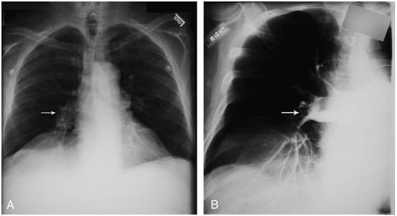
- (A) enlargement of the right interlobar artery (arrow).
- (B) Angiogram confirms the presence of multiple filling defects representing multiple emboli and a distended right interlobar artery (arrow).
History
1958 – Fleischner published a case report of a 39 year old hospitalised man who developed sudden sharp right sided chest pain and dry cough with preceding thrombophlebitis of the right foot and ankle. Fleischner highlighted the prominent right hilar shadow on chest x-ray of the patient and associated this with a large unilateral embolus.
an embolus had lodged in the right pulmonary artery. The mechanical obstruction and vasoconstriction of the peripheral arteries prevented blood flow to the right lung, which appeared oligemic. The right hilar shadow was increased in size, due either to the embolothrombus lodged there or to the pulmonary hypertension, or to both.”
Fleischner, 1958 (in reference to a chest radiograph)
Fleischner sign 2.0
Fleischner sign is most commonly attributed to a dilated central pulmonary artery associated with pulmonary embolus. However, Fleischner lectured and published extensively on pulmonary embolus and pulmonary hypertension between 1941 and 1962. As a result, he has been eponymously affiliated with other CXR changes often observed in pulmonary embolism such as the raised hemidiaphragm with basal atelectasis
Roentgenologically, in addition to the classical infarct, we look for signs of impaired ventilation, such as high position of the diaphragm, diminished ventilatory diaphragmatic excursion and basal atelectasis, often in the form of plate atelectasis. The diminished distance between the right dome of the diaphragm and the horizontal interlobar fissure is a good yardstick for inhibited ventilation. This ventilatory inhibition may occur on one side only or bilaterally.
Fleischner 1958
Respiratory inhibition is frequent and is revealed radiologically by a high position and diminished excursion of the diaphragm, and by basal atelectasis, often in the form of plate atelectasis.
Fleischner 1962
Other Fleischner pulmonary embolism pearls:
- Pulmonary embolism may occur under the disguise of other pulmonary, cardiac, pleural, neurologic or abdominal diseases, and the classical clinical picture is the exception.
- Among the signs and symptoms, chest pain and dyspnea, singly or combined, are the most common early signals. Haemoptysis is comparatively rare…
- If pulmonary infarction is diagnosed by clinical means or roentgenologically, this signifies that there has been an episode of embolism…
- The “triangular shadow” of the infarct is a fiction. Consolidations of any shape may be caused by infarcts. The classical appearance is a consolidation of cushion or hemispherical shape, with its longest extension arranged along a pleural surface…
- Many instances of embolism, probably the majority, occur without infarct formation…
Associated Persons
- Nils Westermark (1892 – 1980) [Westermark sign 1938]
- Aubrey Otis Hampton (1900 – 1955) [Hampton hump 1940]
- Felix George Fleischner (1893 – 1969) [Fleischner sign 1959]
- C. H. Joseph Chang (1929 – 2017) [Chang sign 1965]
- Antonio Palla (1950 – ) [Palla sign 1983]
Alternative names
- Fleischner’s sign
References
- Fleischner FG. Pulmonary embolism. Canadian Medical Association Journal. 1958; 78(9): 653–660.
- Fleischner F. Unilateral pulmonary embolism with increased compensatory circulation through the unoccluded lung. Radiology 1959; 73: 591-597
- Fleischner F. Roentgen diagnosis of pulmonary embolism. Heart Bull. 1961; 10: 104-7.
- Fleischner FG. Pulmonary embolism. Clin Radiol. 1962; 13(3): 169-82.
- Williams JR, Wilcox WC. Pulmonary embolism: roentgenographic and angiographic considerations. AJR Am J Roentgenol. 1963; 89: 333.
- Stein PD, Athanasoulis C, Greenspan RH, et al. Relation of plain chest radiographic findings to pulmonary arterial pressure and arterial blood oxygen levels in patients with acute pulmonary embolism. Am J Cardiol. 1992; 69: 394
- Cadogan M. CXR eponyms in pulmonary embolism. LITFL
eponymictionary
the names behind the name
Doctor in Australia. Keen interest in internal medicine, medical education, and medical history.