![]()
Abdominal CT: abdominal veins
Checking the abdominal and pelvic venous systems
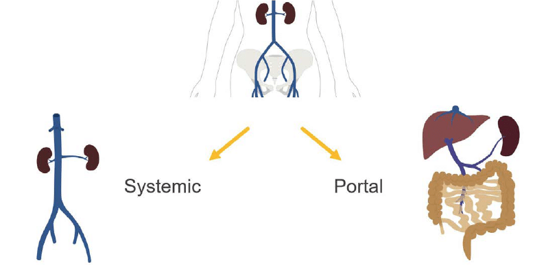
The venous anatomy is divided into systemic and portal venous systems. The key difference between these two systems is the liver.
- Systemic system: Systemic veins drain directly to the heart through the inferior vena cava without being filtered by the liver first, with most of the venous flow coming from the hepatic veins, kidneys, and lower extremities.
- Portal venous system: In the portal venous system, the liver filters the blood returning from the gastrointestinal (GI) tract and spleen before it returns to the heart through the hepatic veins and IVC.
Systemic system
The systemic venous anatomy includes the:
- Inferior vena cava (IVC)
- Renal veins
- Iliac veins
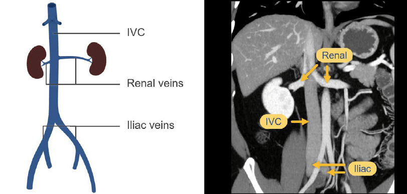
The lower extremity veins receive the contrast-mixed blood a bit slower compared to the kidneys which rapidly light up with contrast. As a result, the renal veins are often brighter than the iliac veins and the lower inferior vena cava, creating a mixing artefact when they join together with the inferior vena cava below the liver.
A mixing artefact can be confused with a clot at first glance, but typically it maintains a layered appearance.
Example:
- Left image: Portal venous phase of the inferior vena cava. The bright contrast mixed blood from the renal veins mixes with the darker blood from the inferior vena cava.
- Right image: Another example of a portal venous phase image of the inferior vena cava but with less mixing artefact, appearing more uniform.
The degree of mixing artefact varies with time. Even a few seconds of difference between scans can change this appearance.

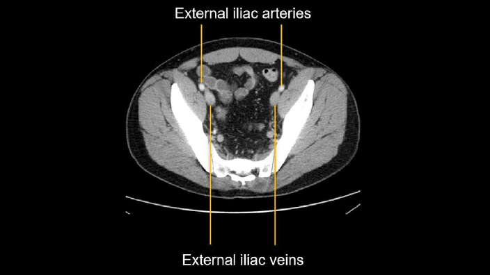
The iliac and common femoral veins are included in abdominopelvic CTs. The brightness of the veins can vary with scan timing, and seeing a mixing artefact with bright or dark blood swirling is fairly common.
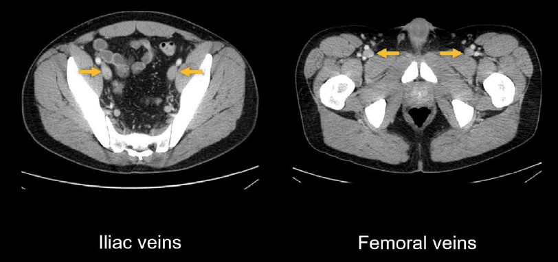
You can differentiate between corresponding external iliac veins and arteries by noticing that the vein is posterior to (i.e., behind) the artery, less bright, and larger.
Portal venous system
Blood returning from the structures of the gastrointestinal tract and spleen is first carried to the liver through the portal venous system. This nutrient-rich blood is then filtered and processed by the liver before joining the systemic circulation through the hepatic veins.
The key portal venous anatomy on CT starts with the mesenteric veins – specifically the superior mesenteric vein (SMV) which joins with the splenic vein to form the portal vein. This relationship is often best seen on coronal images.
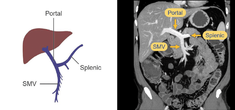
The post-processed CT image below allows you to see thicker segments of the vasculature in a single image and helps you view the relationship of the superior mesenteric, splenic, and portal veins without having to scroll through multiple images. The point where these veins converge in the upper abdomen is called the portosplenic confluence, an important anatomic landmark to keep in mind.

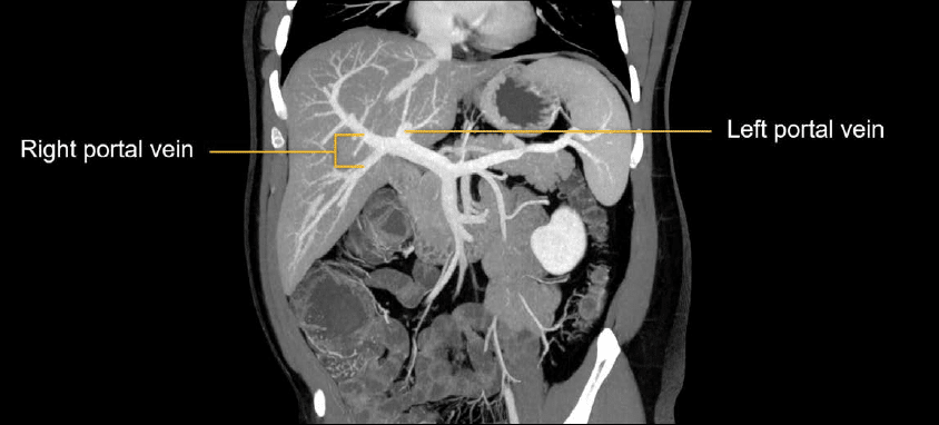
Let’s take a closer look at the portal vein as it enters the liver. The main portal vein splits into right and left branches as it enters the liver and delivers nutrient rich blood from the GI tract and spleen.
After the portal blood is filtered and processed by the liver, it returns to the systemic circulation through the hepatic veins, into the inferior vena cava, and finally the right atrium.

Knowledge iteration
To solidify the core anatomy, scroll through this set of images on a PACS viewer. You can get a sense of how to efficiently review this important venous anatomy to reinforce these key relationships. Try scrolling through the coronal images with the axial next to them for reference
- Start with the superior mesenteric vein, which joins with the splenic vein at the portal splenic confluence and supplies the portal vein.
- The portal vein then heads to the liver and splits into the major right and left branches.
- Next, head down to the pelvis for the common femoral and iliac veins. These often have mixing artefact on portal venous phase CTs.
- Following the common femoral and iliac veins up into the pelvis, you will reach the inferior vena cava, renal veins, intrahepatic segment of the inferior vena cava, and the right atrium.
- Coronal images nicely layout this anatomy, where you can see the renal veins emptying into the IVC with mixing artefact from the unenhanced pelvic drainage, heading up to the right atrium.
This is an edited excerpt from the Medmastery course Abdominal CT Essentials by Michael P. Hartung, MD. Acknowledgement and attribution to Medmastery for providing course transcripts.
- Hartung MP. Abdominal CT: Common Pathologies. Medmastery
- Hartung MP. Abdominal CT: Essentials. Medmastery
- Hartung MP. Abdomen CT: Trauma. Medmastery
References
- Top 100 CT scan quiz. LITFL
Radiology Library: Abdominal CT: Imaging important abdominal structures
- Hartung MP. Abdominal CT: Checking the abdominal arteries
- Hartung MP. Abdominal CT: Checking the abdominal veins
- Hartung MP. Abdominal CT: Screening the lymph nodes
- Hartung MP. Abdominal CT: Evaluating the body wall
- Hartung MP. Abdominal CT: Evaluating the bones
- Hartung MP. Abdominal CT: Reporting checklist
Abdominal CT interpretation

Assistant Professor of Abdominal Imaging and Intervention at the University of Wisconsin Madison School of Medicine and Public Health. Interests include resident and medical student education, incorporating the latest technology for teaching radiology. I am also active as a volunteer teleradiologist for hospitals in Peru and Kenya. | Medmastery | Radiopaedia | Website | Twitter | LinkedIn | Scopus

MBChB (hons), BMedSci - University of Edinburgh. Living the good life in emergency medicine down under. Interested in medical imaging and physiology. Love hiking, cycling and the great outdoors.