![]()
Abdominal CT: abdominal arteries
Checking the abdominal arteries
On a standard portal venous phase computed tomography (CT) series, you will get a balanced view of the vasculature that includes both major arteries and veins. On the other hand, an examination such as CT angiography obtains images earlier after contrast which allows you to see even small arterial branches.
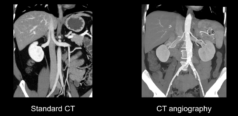
At times, it can be tough to confidently evaluate the vasculature. Often, CT images have some blurring due to patient motion, and the vessels can be affected by mixing artefact. Mixing artefact occurs when blood with different amounts of contrast mixes together and creates lines or swirls in the vessels. However, these limitations don’t prevent us from trying!
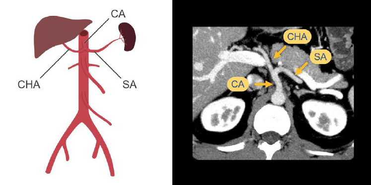
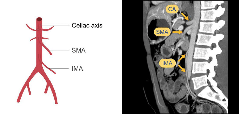
Coeliac axis
Starting with the arterial anatomy, we can trace the abdominal aorta to its first major branch – the coeliac axis (CA). The coeliac axis splits into the common hepatic artery (CHA) and the splenic artery (SA). You can often follow these into their distal branches, but such a detailed review is usually reserved for a dedicated CT angiography exam.
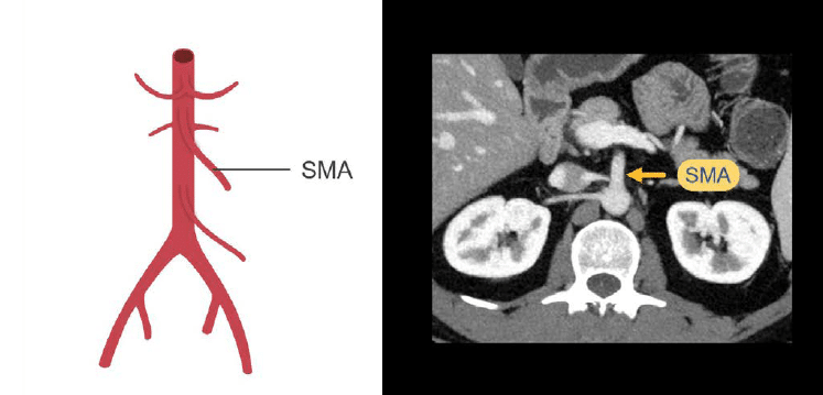
Superior mesenteric artery
The next major branch of the abdominal aorta as we head toward the feet is the superior mesenteric artery (SMA) which supplies the small bowel and the right colon.
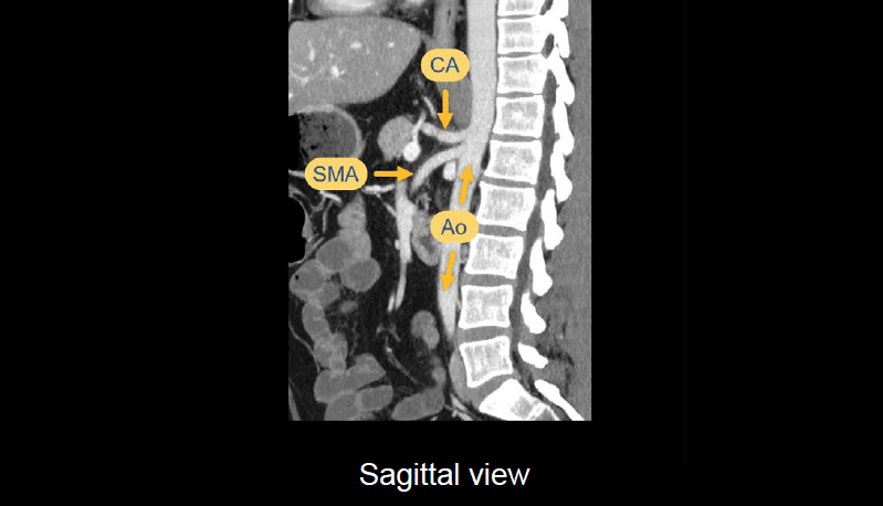
If we look at the sagittal view, we can see the long axis of the abdominal aorta (Ao) and the close proximity of the origins of the celiac axis, which curves slightly upwards, and the superior mesenteric artery, which curves downwards to supply the small bowel. This is a helpful view to keep in mind for vascular evaluation.
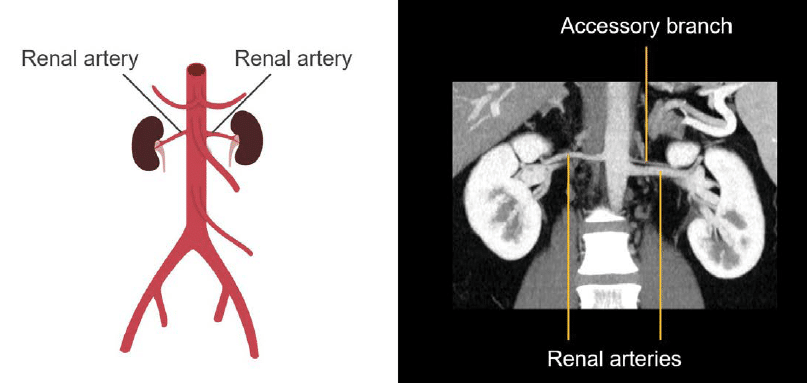
Renal arteries
The renal arteries are the next major branch to evaluate. In the coronal image below, we can see the right and left renal arteries as well as a smaller accessory branch on the left. Accessory renal artery branches are common and can be seen on both sides in this case.
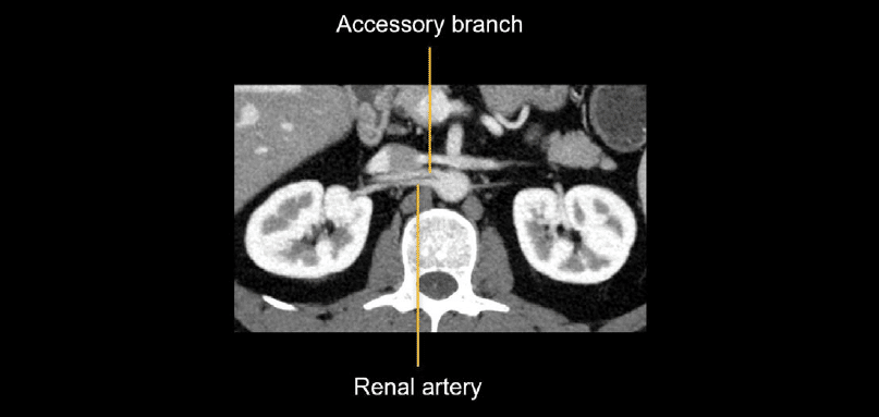
The axial image below allows you to see both the renal artery and an accessory branch on the right side, which helps us to complete the picture of the arterial anatomy.
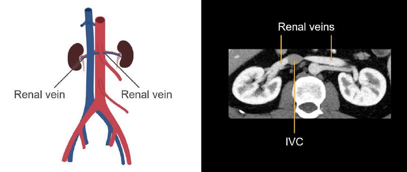
It is helpful to keep in mind the relationship between the renal veins and arteries, as you will often see them on the same images. Below we can also see portions of the renal veins coursing in front of the renal arteries and joining with the inferior vena cava (IVC).
Inferior mesenteric artery
The inferior mesenteric artery (IMA) is a small branch of the mid abdominal aorta that supplies the left colon. It is a good deal smaller than the SMA, and for this reason, it can be hard to see, unless you know where to look.

On the sagittal image below, we can see the relative spacing of the celiac axis and superior and inferior mesenteric arteries as they branch from the abdominal aorta. When looking for the inferior mesenteric artery, locate an anterior branch that is heading down toward the pelvis starting about halfway down the abdominal aorta.
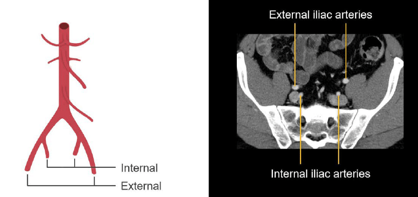
Common iliac arteries
Finally, as we reach the end of the aorta, it splits into the common iliac arteries at the aortic bifurcation. The common iliac arteries then split into the internal and external iliac arteries supplying the pelvis before descending into the upper thighs as the femoral arteries.

Knowledge iteration
To solidify the core anatomy, scroll through this set of images on a PACS viewer. You can get a sense of how to efficiently review this important vasculature. Scroll through the axial images with the coronal images next to them.
- Start with the distal thoracic aorta and follow it as it passes through the diaphragm to find the first major branch – the coeliac axis. The celiac axis splits into the common hepatic artery toward the patient’s right, which goes on to supply the liver, and the splenic artery to the left, which follows a winding course to the spleen.
- The next major branch is the SMA, which splits into several branches supplying the small bowel and the right colon.
- By going back up to the SMA origin, you can see the renal arteries supplying the right and left kidneys.
- The last major branch of the abdominal aorta is the rather small inferior mesenteric artery, which supplies the left colon.
- Finally, the aorta splits at the aortic bifurcation into the common iliac arteries, internal and external iliac arteries, and then into the common femoral arteries in the upper thighs.
This is an edited excerpt from the Medmastery course Abdominal CT Essentials by Michael P. Hartung, MD. Acknowledgement and attribution to Medmastery for providing course transcripts.
- Hartung MP. Abdominal CT: Common Pathologies. Medmastery
- Hartung MP. Abdominal CT: Essentials. Medmastery
- Hartung MP. Abdomen CT: Trauma. Medmastery
References
- Top 100 CT scan quiz. LITFL
Radiology Library: Abdominal CT: Imaging important abdominal structures
- Hartung MP. Abdominal CT: Checking the abdominal arteries
- Hartung MP. Abdominal CT: Checking the abdominal veins
- Hartung MP. Abdominal CT: Screening the lymph nodes
- Hartung MP. Abdominal CT: Evaluating the body wall
- Hartung MP. Abdominal CT: Evaluating the bones
- Hartung MP. Abdominal CT: Reporting checklist
Abdominal CT interpretation

Assistant Professor of Abdominal Imaging and Intervention at the University of Wisconsin Madison School of Medicine and Public Health. Interests include resident and medical student education, incorporating the latest technology for teaching radiology. I am also active as a volunteer teleradiologist for hospitals in Peru and Kenya. | Medmastery | Radiopaedia | Website | Twitter | LinkedIn | Scopus

MBChB (hons), BMedSci - University of Edinburgh. Living the good life in emergency medicine down under. Interested in medical imaging and physiology. Love hiking, cycling and the great outdoors.