![]()
Abdominal CT: bones
Evaluating the bones
The bones are often one of the last items on the reporting checklist for abdominal CT, but they still deserve our careful attention.
When evaluating the bones, be sure to switch to the bone window to make the job easier. Bone windows will help you see the detail of the bone structure including the outer, dense cortex and the inner medullary cavity
Spine
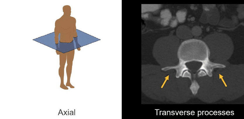
It is hard to evaluate the vertebral body height on axial images, but we can look at the posterior elements and transverse processes while benefitting from the symmetry in this view.
Sagittal is the best view for reviewing the vertebral body height and alignment as you can see and compare all of the vertebrae in a single image. In the image shown next, we can see how the lumbar and lower thoracic vertebral bodies stack on top of each other, and we can also see the spinous processes posteriorly.

Hips and pelvis
The coronal view is particularly helpful for evaluating the symmetry of the hips and pelvis. It will help you see the length of the sacroiliac joints and sacrum, as well as compare both hip joints with each other.

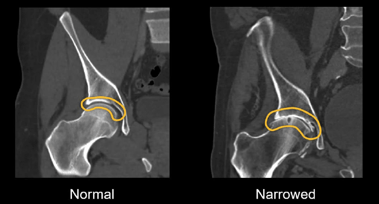
When evaluating the hip joints, it is common to see a narrowing of the space between the head of the femur and the acetabulum related to arthritis.
As CT x-rays can’t penetrate the metal used for a hip replacement, you may also see a metallic streak artefact or bright metal from total hip replacements. In the case featured next, this appears as black and white streaks in the image.

Degenerative disease
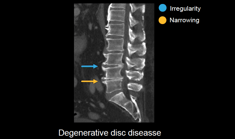
Similar to degenerative disease of the hips, in the spine, it is common to see narrowing and irregularity of the vertebral body end plates, particularly in older patients. This is broadly referred to as degenerative disc disease. Detailed evaluation of this is typically reserved for magnetic resonance imaging (MRI).
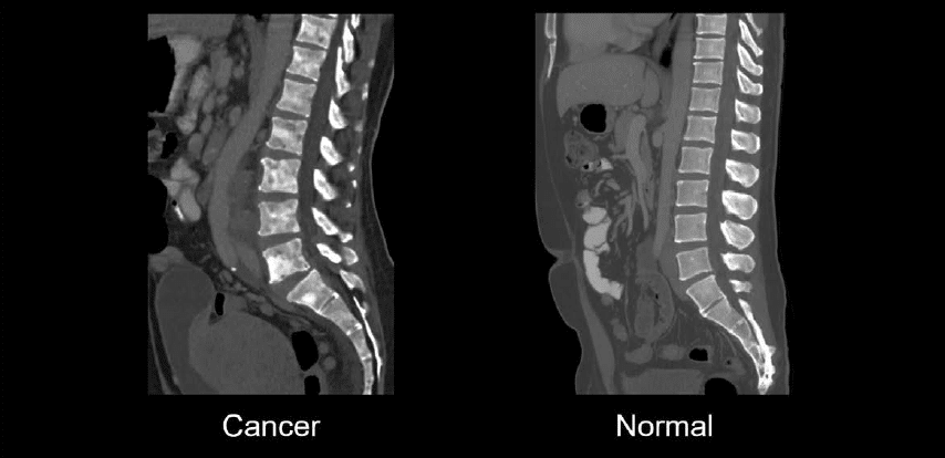
Cancer
When cancer spreads to the bone, it can cause areas of increased density (which appear brighter) or decreased density.
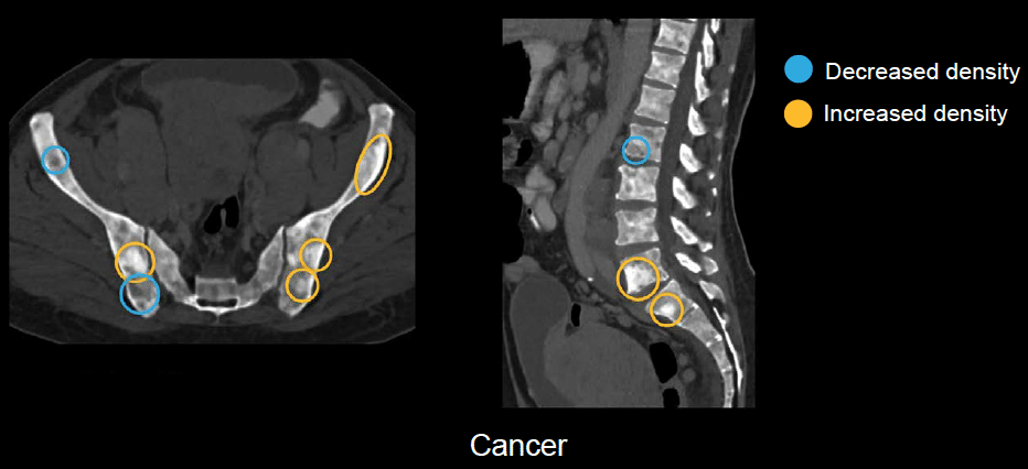
In our next case featuring a patient with metastatic prostate cancer, the entire spine and pelvis are involved with the tumour, making the bones look patchy and irregular with alternating areas of varying brightness compared to the relatively uniform and smooth look of the normal spine.
Knowledge iteration
Try out an evaluation of the bones by reviewing this set of images with an online PACS viewer
- Bone evaluation starts with bone window and axial view. Look for the:
- lower ribs
- lower thoracic vertebral bodies
- lumbar spine
- sacrum
- sacroiliac joints
- iliac bones
- acetabulum
- hip joints
- superior pubic ramus
- pubic body
- inferior pubic ramus
- Switching to coronal, you can get an intuitive view of the following:
- proximal femur
- hip joints
- sacroiliac joints
- sacrum
- The sagittal view lays out the long axis of the spine, evaluate:
- vertebral body height and alignment
- posterior elements of the vertebrae including the pedicles and spinous processes
- The sagittal view provides this excellent view of the lower ribs.
- Most importantly, the sagittal view lays out the long axis of the spine, to evaluate the:
- alignment
- end plates
- posterior elements of the vertebrae including the spinous processes and articular pillars
This is an edited excerpt from the Medmastery course Abdominal CT Essentials by Michael P. Hartung, MD. Acknowledgement and attribution to Medmastery for providing course transcripts.
- Hartung MP. Abdominal CT: Common Pathologies. Medmastery
- Hartung MP. Abdominal CT: Essentials. Medmastery
- Hartung MP. Abdomen CT: Trauma. Medmastery
References
- Top 100 CT scan quiz. LITFL
Radiology Library: Abdominal CT: Imaging important abdominal structures
- Hartung MP. Abdominal CT: Checking the abdominal arteries
- Hartung MP. Abdominal CT: Checking the abdominal veins
- Hartung MP. Abdominal CT: Screening the lymph nodes
- Hartung MP. Abdominal CT: Evaluating the body wall
- Hartung MP. Abdominal CT: Evaluating the bones
- Hartung MP. Abdominal CT: Reporting checklist
Abdominal CT interpretation

Assistant Professor of Abdominal Imaging and Intervention at the University of Wisconsin Madison School of Medicine and Public Health. Interests include resident and medical student education, incorporating the latest technology for teaching radiology. I am also active as a volunteer teleradiologist for hospitals in Peru and Kenya. | Medmastery | Radiopaedia | Website | Twitter | LinkedIn | Scopus

MBChB (hons), BMedSci - University of Edinburgh. Living the good life in emergency medicine down under. Interested in medical imaging and physiology. Love hiking, cycling and the great outdoors.