
![]()
Abdominal CT: lymph nodes
Screening the lymph nodes
Evaluating for abnormal or enlarged lymph nodes is an essential part of any abdominal CT, particularly when staging cancer. Lymph node territories are named based on their anatomic location (i.e., what organ or structure they are next to or what major blood vessel they are following).
Upper abdomen
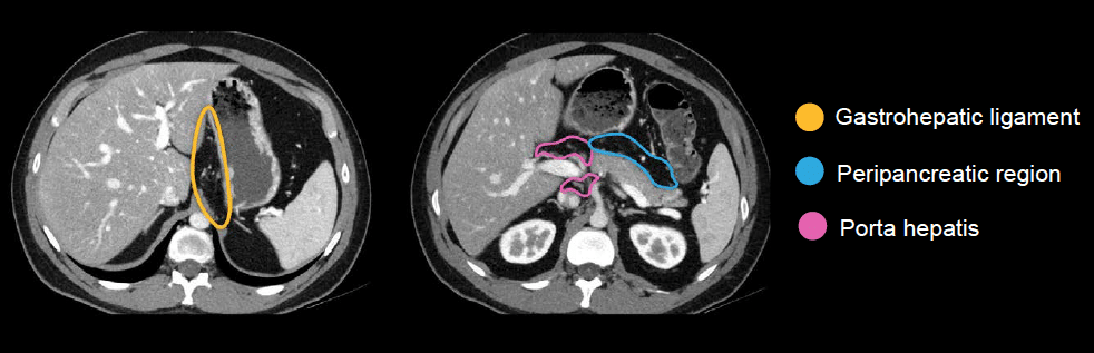
Starting in the upper abdomen, we check three locations:
- Gastrohepatic ligament region (between the stomach and liver)
- Porta hepatis region (around the vessels entering the liver)
- Peripancreatic region (surrounding the pancreas)
In the small bowel mesentery
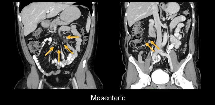
There are lymph nodes that extend out into the small bowel mesentery as well. Normally, these look like small, round dots and are often best seen on coronal images. These are broadly referred to as mesenteric lymph nodes and, more specifically, ileocolic lymph nodes in the right lower quadrant heading toward the cecum and terminal ileum.
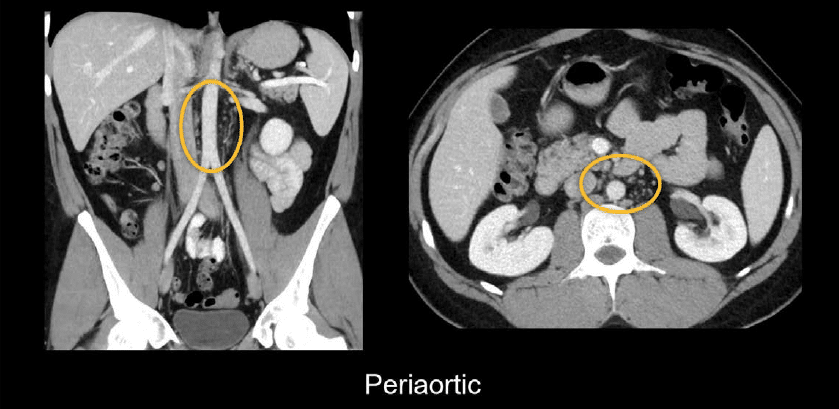
Around the abdominal aorta
Next, we can broadly consider the area surrounding the abdominal aorta as periaortic or retroperitoneal. This distribution is also nicely seen on coronal images.
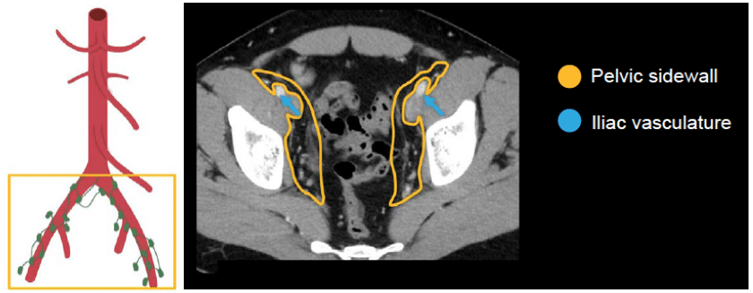
In the pelvic region
As we head into the pelvis, we can follow the distribution of the iliac vasculature for the accompanying lymph nodes. The pelvic sidewall broadly refers to the space in the pelvis that surrounds the vasculature and includes these lymph nodes.
The inguinal region is a common place to see lymph nodes. Inguinal lymph nodes are usually relatively small and symmetric.
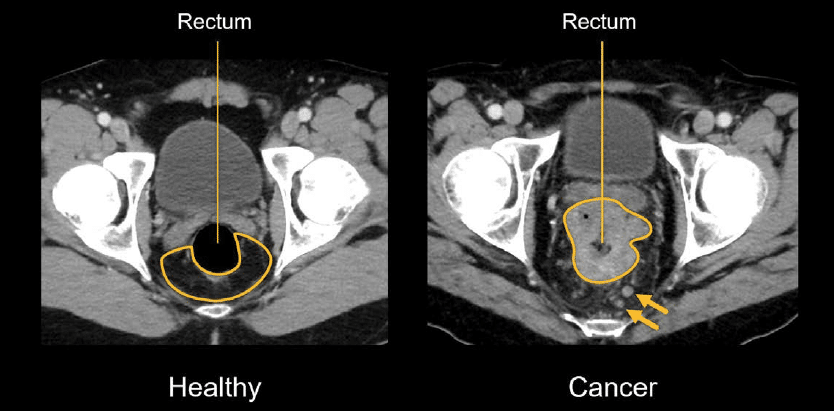
In a healthy individual, it is unlikely you would see lymph nodes in the space around the rectum. However, this is a common site for cancer to spread and in this scenario, we may find enlarged or abnormal lymph nodes, particularly with rectal and prostate cancers.
Assessing enlarged lymph nodes
There are a variety of inflammatory, infectious, and neoplastic causes that result in enlarged lymph nodes. The clinical context and distribution of the lymph nodes will help to determine the likelihood that enlarged lymph nodes are related to cancer.
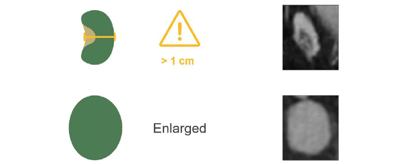
Lymph nodes are generally measured along their short axis, and typically a size greater than 1 cm is considered abnormal. Enlarged lymph nodes become increasingly rounded and lose their characteristic fatty hilum.
On CT, enlarged lymph nodes usually look like rounded nodules with soft tissue density.
Examples
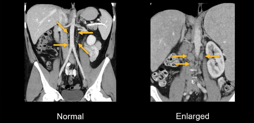
In the coronal example of the periaortic region below, we can see multiple enlarged lymph nodes around the aorta
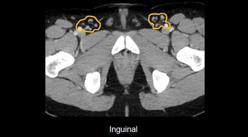
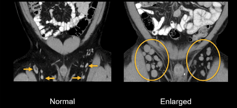
When compared to normal lymph nodes, the patient below has numerous enlarged inguinal lymph nodes.

In the coronal images from the same patient, you can really see how many enlarged inguinal lymph nodes there are on both sides compared to the normal example. These lymph nodes are large, rounded, and have lost their normal fatty hilum.
Knowledge iteration
To solidify your knowledge, scroll through this set of images on a PACS viewer. You can put all of these important nodal territories together into a cohesive search pattern starting with the upper abdomen and working your way down.
- Between the stomach and liver, look for the gastrohepatic ligament, which is an important location to watch when staging esophageal and gastric cancers.
- Around the celiac axis, you can see several small lymph nodes and the celiac ganglia; this area is important for screening and staging pancreatic cancer.
- Look around the liver hilum (i.e., lymph nodes at the porta hepatis) and between the portal vein and inferior vena cava (i.e., portacaval nodes). We can also screen in the area around the pancreas (i.e., peripancreatic nodes).
- Before continuing with the aorta, you can follow the folds of the mesentery out and evaluate for enlarged mesenteric and ileocolic lymph nodes.
- Next, come back up the abdomen and run the aorta all the way down. This area is broadly referred to as periaortic. Between the aorta and inferior vena cava, we can be more specific and call these aortocaval nodes.
- By heading down to the external and internal iliac arteries, you can run the vessels and sidewall, around the rectum for the mesorectum, and finally into the inguinal regions.
This is an edited excerpt from the Medmastery course Abdominal CT Essentials by Michael P. Hartung, MD. Acknowledgement and attribution to Medmastery for providing course transcripts.
- Hartung MP. Abdominal CT: Common Pathologies. Medmastery
- Hartung MP. Abdominal CT: Essentials. Medmastery
- Hartung MP. Abdomen CT: Trauma. Medmastery
References
- Top 100 CT scan quiz. LITFL
Radiology Library: Abdominal CT: Imaging important abdominal structures
- Hartung MP. Abdominal CT: Checking the abdominal arteries
- Hartung MP. Abdominal CT: Checking the abdominal veins
- Hartung MP. Abdominal CT: Screening the lymph nodes
- Hartung MP. Abdominal CT: Evaluating the body wall
- Hartung MP. Abdominal CT: Evaluating the bones
- Hartung MP. Abdominal CT: Reporting checklist
Abdominal CT interpretation

Assistant Professor of Abdominal Imaging and Intervention at the University of Wisconsin Madison School of Medicine and Public Health. Interests include resident and medical student education, incorporating the latest technology for teaching radiology. I am also active as a volunteer teleradiologist for hospitals in Peru and Kenya. | Medmastery | Radiopaedia | Website | Twitter | LinkedIn | Scopus

MBChB (hons), BMedSci - University of Edinburgh. Living the good life in emergency medicine down under. Interested in medical imaging and physiology. Love hiking, cycling and the great outdoors.
