Befuddling Pupillary Asymmetry
aka Ophthalmology Befuddler 001
A medical student on your team asks you to review an 81 year-old female who speaks little English. She was BIBA to the ED following a fall. Her nursing home transfer sheet says that the fall was witnessed: she tripped and there was no loss of consciousness. The student is concerned that the patient’s right pupil is fixed and slightly dilated in the presence of facial abrasions. Facial views have been ordered.
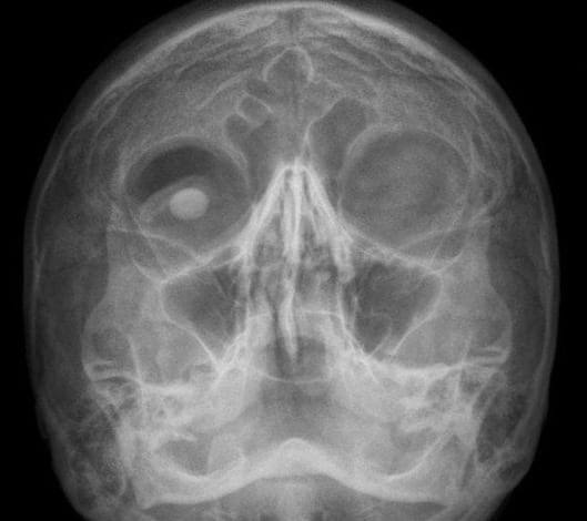
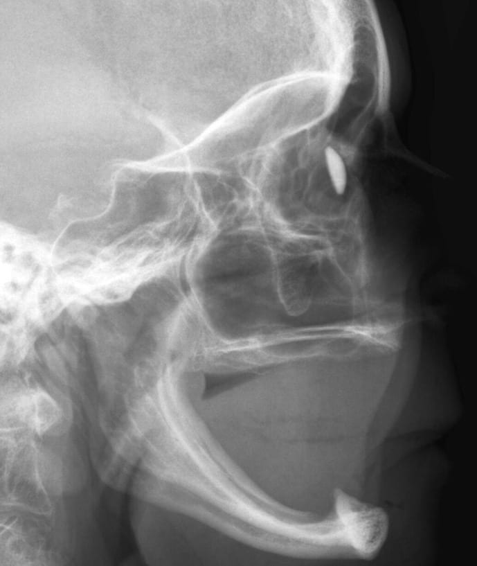
Before you go to see the patient you check to see if the radiographs are available to view. This is what you see:


Q1. What is the abnormality?
Answer and interpretation
The patient has an right-sided ocular prosthesis – an artificial eye! – and no teeth…

Check out this reference for more radiographic images of head and neck devices and prostheses:
Hunter, T., Yoshino, M., Dzioba, R., Light, R., & Berger, W. (2004). Medical Devices of the Head, Neck, and Spine Radiographics, 24 (1), 257-285 DOI: 10.1148/rg.241035185
Ever wondered how an eye prosthesis is made?
Q2. What is anisocoria?
Answer and interpretation
Anisocoria simply means that there is inequality in the size of the pupils.
Q3. What are the possible causes of anisocoria?
Answer and interpretation
There are two possible causes:
- one pupil is abnormally small or constricted (miotic)
OR - one pupil is abnormally large or dilated (mydriatic)
Easy, eh.
Actually there is a third option: normal physiological anisocoria.
Learn more:
- The causes of small pupils (miosis) are considered in Neurological Mind-boggler 002.
- GSA HEMS — OXY’s LOG – ‘Anisocoria and Stardust’…
Q4. What are the causes of a fixed dilated pupil in a conscious patient?
Answer and interpretation
Pharmacologic blockade is the most common cause of a fixed dilated pupil in an otherwise normal healthy patient.
A fixed dilated pupil in an awake patient is NOT due to herniation.
A single fixed dilated (mydriatic) pupil can be caused by:
Pharmacological blockade: typically topical mydriatic drugs used to facilitate ophthalomological examinations.
- anticholinergic drugs: e.g. atropine, cyclopentolate and tropicamide
- alpha1-agonists: phenylephrine.
Oculomotor nerve palsy: (3rd cranial nerve)
- parasympathetic nerves are in the superficial parts of the nerve, so tend to be more vulnerable to compressive lesions and spared by vascular lesions (e.g. diabetes mellitus).
- If an acute third nerve palsy is accompanied by pupillary mydriasis an aneurysm arising from the posterior communicating artery must be excluded.
[See a Spider called Willis for an easy way to remember the components of the Circle of Willis and its relations.]
Other:
- Holmes-Adie pupil (tonic phase)
- post-traumatic iridocyclitis (e.g. direct facial trauma)
- acute closed-angle glaucoma
- physiological anisocoria
- ocular prosthesis – the normal pupil may be relatively constricted due to ambient light.
Now, consider a patient with these findings on clinical examination:
Q5. What is the abnormality?
Answer and interpretation
A left-sided Holmes-Adie pupil.
Q6. Describe the features of this abnormality.
Answer and interpretation
A Holmes-Adie pupil is a pupillary abnormality of uncertain etiology associated with postganglionic parasympathetic denervation. It is usually found in an otherwise healthy young woman.
- The acutely large pupil diminishes in size over months to years. It is sluggish or non-reactive to light and has a slow (tonic) near response.
- At the slit-lamp, slow vermiform contractions of the iris may be seen.
- The diagnosis may be clinched by demonstrating the presence of hypersensitivity to weak miotic eye drops (constriction with 0.05-0.1% pilocarpine eye drops) that have little effect on normal pupils.
Here is another visual demonstration of the features of the Holmes-Adie pupil:
The attribution of this eponym has a fairly convoluted history — but is in part named for a brilliant Irishman renowned for his attention to detail and refusal to bend facts to fit a hypothesis — it has nothing to do with the fictional detective based (Smith, Bell and the art of observation).
References
- Bhidayasiri R, Waters MF, Giza CC. Neurological differential diagnosis: a prioritized approach, Blackwell Publishing 2005.
- Patten J. Neurological differential diagnosis (2nd edition), Springer-Verlag 1996.
- Sir Gordon Morgan Holmes (1876-1965)
- William John Adie (1886-1935)

OPHTHALMOLOGY BEFUDDLER
Chris is an Intensivist and ECMO specialist at The Alfred ICU, where he is Deputy Director (Education). He is a Clinical Adjunct Associate Professor at Monash University, the Lead for the Clinician Educator Incubator programme, and a CICM First Part Examiner.
He is an internationally recognised Clinician Educator with a passion for helping clinicians learn and for improving the clinical performance of individuals and collectives. He was one of the founders of the FOAM movement (Free Open-Access Medical education) has been recognised for his contributions to education with awards from ANZICS, ANZAHPE, and ACEM.
His one great achievement is being the father of three amazing children.
On Bluesky, he is @precordialthump.bsky.social and on the site that Elon has screwed up, he is @precordialthump.
| INTENSIVE | RAGE | Resuscitology | SMACC
