![]()
Jean-Gaspard Blaise Goyrand
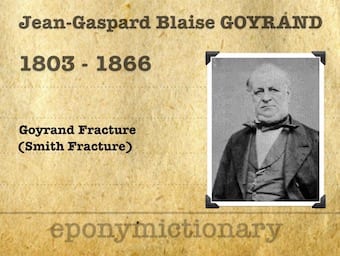
Jean-Gaspard Blaise Goyrand (1803 – 1866) was a French surgeon. Eponym: Goyrand Fracture (France - 1832) Smith fracture, wrist fracture

![]()
Jean-Gaspard Blaise Goyrand (1803 – 1866) was a French surgeon. Eponym: Goyrand Fracture (France - 1832) Smith fracture, wrist fracture
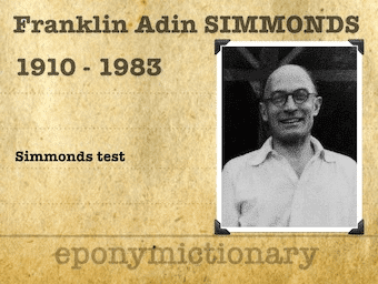
Franklin Adin 'Sam' Simmonds (1910–1983) British orthopaedic surgeon. Simmonds-Thompson Test for Achilles tendon rupture with TC Thompson (1902-1986)

T. Campbell Thompson (1902 – 1986) American Orthopedic Surgeon. Simmonds-Thompson Test for Achilles tendon rupture with Sam Simmonds (1910–1983)
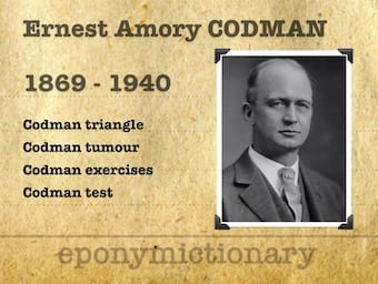
Ernest Amory Codman (1869-1940) was an American surgeon. Founder of an 'end results system' to track the outcomes of patient treatments.

Plaster tips and tricks with Dan Smith; Splint immobilisation, full casts; bi-valves; and plaster removal techniques

Hoffmann-Tinel sign is paresthesia in the distal cutaneous distribution of an injured peripheral nerve evoked by tapping on the nerve more proximally.
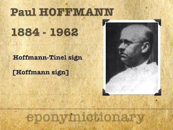
Paul Hoffmann (1884-1962) was a German physiologist and physician. Known for describing Hoffmann-Tinel sign for assessment of nerve regeneration and success of nerve sutures.
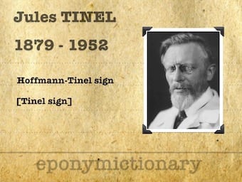
Jules Tinel (1879 – 1952) was a French neurologist. Eponymously affiliated with Tinel's sign in the diagnsois of carpal tunnel syndrome
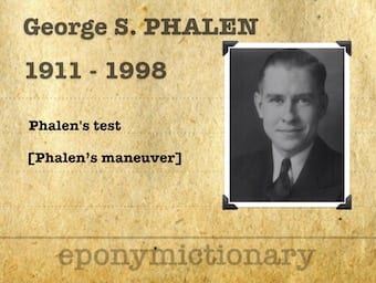
George S. Phalen (1911 – 1998) was an American Orthopedic Surgeon. Phalen defined our understanding of carpal tunnel syndrome aetiology, assessment and management
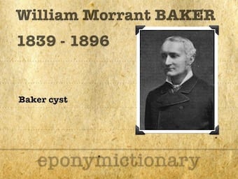
William Morrant Baker (1839 – 1896) was a British General Surgeon. Eponymously affiliated with the Baker's cyst and Baker's cannula, a flexible tracheal cannula
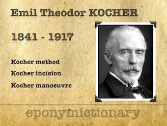
Emil Theodor Kocher (1841 – 1917) was a Swiss Surgeon. Nobel Prize in Physiology or Medicine 1909. 5,000 thyroid excisions. Shoulder reduction

Johan Henning Waldenström (1877-1972) Swedish Orthopaedic surgeon. Specialised in pediatric bone/joint tuberculosis Legg-Calvé-Perthes disease 1909