![]()
Network Five: Practice Changes In Paediatric Wheeze Management
Network Five Emergency Medicine Journal Club Episode 30 reviewing updates on paediatric wheeze management with paediatric respiratory and sleep specialist Dr Chetan Pandit!

![]()
Network Five Emergency Medicine Journal Club Episode 30 reviewing updates on paediatric wheeze management with paediatric respiratory and sleep specialist Dr Chetan Pandit!
Löffler (Loeffler) syndrome is a transient, self-limiting, and benign pulmonary eosinophilia, characterised by pulmonary opacities on X-ray, elevated blood eosinophils and an acute onset of potential symptoms of mainly cough and dyspnoea.

Wilhelm Löffler (1887 – 1972) was a Swiss physician. Löffler is eponymously associated with two clinical manifestations of eosinophilia which he described: transient pulmonary infiltrates with eosinophilia (Löffler syndrome, 1932) and endocarditis parietalis fibroplastica (Löffler endocarditis, 1936).
Network Five Emergency Medicine Case 1 discussing an interesting case of a patient who presents with chest pain and pre-syncope.
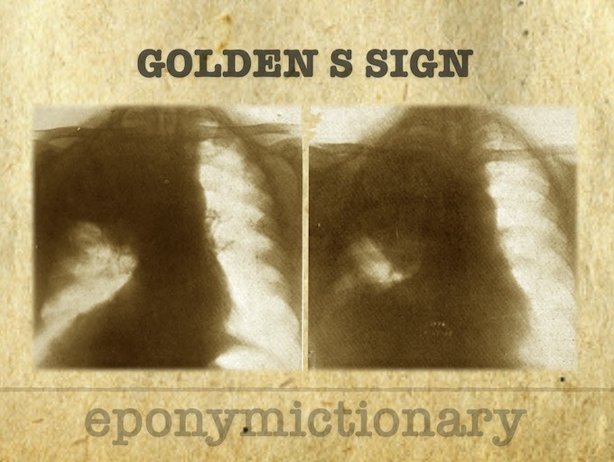
Golden S sign. Radiological sign which should raise suspicion of bronchial carcinoma. Rss Golden first described a characteristic reverse S-shaped shadow in the right upper lobe in 1925
Julian Dobranowski video helps you classify and identify different types of atelectasis on Chest X-ray. Atelectasis defined as reduced inflation of all, or part, of the lung.
Julian Dobranowski and Medmastery video to help you search for and identify a pneumothorax on CXR.
Julian Dobranowsk Medmastery help identify the features of cardiac failure on the chest X-ray. Evaluation of the three stages of failure
Medmastery video helps identify the various radiological presentations of pneumonia such as lobar, interstitial , bronchopneumonia and special considerations in immunocompromised patients
Biography Medical Eponyms Lovibond angle (profile sign) [1938] In 1938, Lovibond was among the first to offer a criteria for the diagnosis of finger clubbing. He defined the ‘profile sign’ of the thumb (Lovibond’s angle), as the angle made by…
Network Five Emergency Medicine Journal Club Episode 11 - Pulmonary embolism with PERT teams, YEARS and the RELAX-PE studies

Professor Abraham Leo Schamroth (1924-1988) was a South African cardiologist.