


ECG Case 003
Middle-aged diabetic patient presenting with shortness of breath. Clinical evidence of pulmonary oedema.
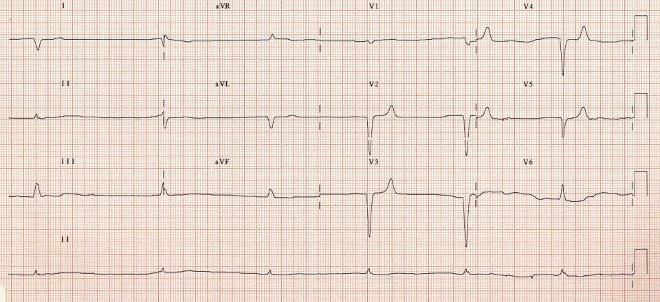
Describe and interpret this ECG
ECG ANSWER and INTERPRETATION
Main Abnormal Findings
- Severe bradycardia of 36 bpm
- Rhythm is difficult to ascertain — appears irregular (?slow AF) although there are some small-voltage P waves seen in V1-2
- Broad QRS complexes with an atypical LBBB morphology
- Subtle symmetrical peaking (“tenting”) of the T waves in V2-5
Diagnosis
The combination of bradycardia, flattening and loss of P waves, QRS broadening and T wave abnormalities is highly suspicious for severe hyperkalaemia. This patient had a potassium of 8.0 in the context of anuric renal failure.
CLINICAL PEARLS
When you see the combination of…
- Bradycardia
- Blocks — e.g. AV block, bundle branch blocks
- Bizarre QRS complexes
…. think hyperkalaemia!
The push-pull effect
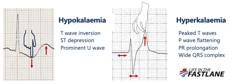
- Hypokalaemia creates the illusion that the T wave is “pushed down”, with resultant T-wave flattening/inversion, ST depression, and prominent U waves
- In hyperkalaemia, the T wave is “pulled upwards”, creating tall “tented” T waves, and stretching the remainder of the ECG to cause P wave flattening, PR prolongation, and QRS widening
References
Further Reading
- Wiesbauer F, Kühn P. ECG Mastery: Yellow Belt online course. Understand ECG basics. Medmastery
- Wiesbauer F, Kühn P. ECG Mastery: Blue Belt online course: Become an ECG expert. Medmastery
- Kühn P, Houghton A. ECG Mastery: Black Belt Workshop. Advanced ECG interpretation. Medmastery
- Rawshani A. Clinical ECG Interpretation ECG Waves
- Smith SW. Dr Smith’s ECG blog.
- Wiesbauer F. Little Black Book of ECG Secrets. Medmastery PDF
TOP 100 ECG Series

Emergency Physician in Prehospital and Retrieval Medicine in Sydney, Australia. He has a passion for ECG interpretation and medical education | ECG Library |

MBBS FACEM DDU (Emergency) CCPU. Emergency Physician in Melbourne, Australia. Co-Ultrasound Lead for Emergency Medicine at The Alfred Hospital. Special interests in diagnostic and procedural ultrasound, medical education, and ECG interpretation. Editor of the LITFL ECG Library.
Seeing that this is a diagnosis of Hyperkalemia. Would it still be appropriate to treat the patient per ACLS for Symptomatic Bradycardia (Atropine, Pacing, Epi or dopamine)? I don’t think many health care professionals would pick up on Hyper K right away but rather treat the patient as they are presenting and fix the rate issue. Am I right? Also, would sodium bicarbonate be appropriate in this situation with a good history and 12 ECG diagnosis?
I think a treatment according to ACLS protocol would be totally appropriate but treating the HyperK will be the reversible cause and therefore be more effective. The main issue being re-&/depolarization issues due to shifted electrolyte gradients: giving i.v.-Calcium is going to increase the heart rate aswell as bridge your patient to elimination (most likely hemodialysis in regards to prior medical history ofc.). Atropine will most likely not have a significant effect on this patient. Epi will though – as it also shifts potassium inside the cells (beta-agonistic adrenergic effect) + increases the heart rate (↑sympathetic tone).