


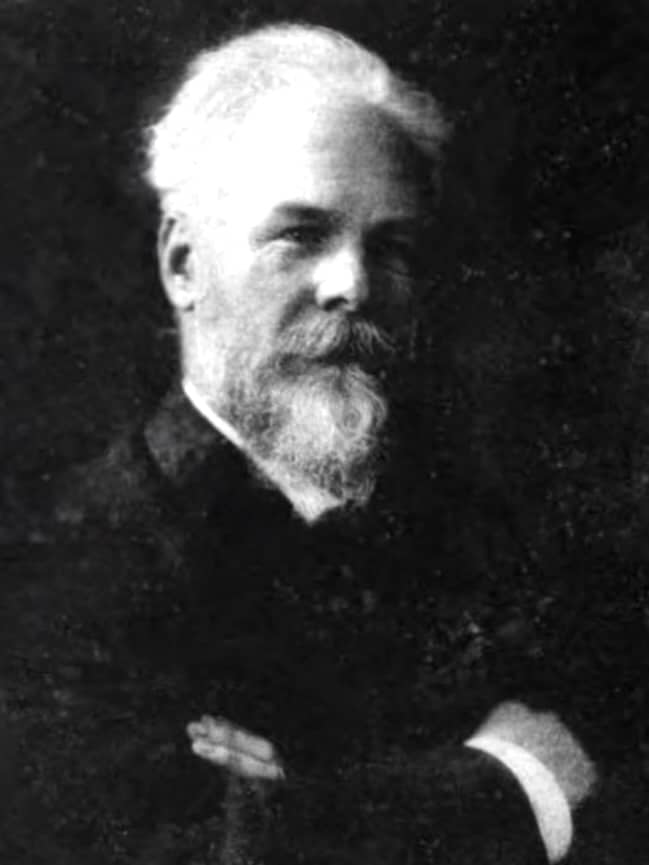
Gustav Killian (1860-1921) was a German laryngologist and rhinologist
Killian helped establish modern endoscopic surgery of the airway and upper gastrointestinal tract. He is best remembered for developing direct bronchoscopy into a practical diagnostic and therapeutic procedure and is regarded as the father of bronchoscopy.
In 1897, Killian successfully passed a rigid oesophagoscope to remove a foreign body from the right main bronchus under direct vision. He went on to develop new instruments and establish Freiburg as an international centre for bronchoscopic training. His introduction of suspension laryngoscopy provided stable exposure of the larynx and allow both hands free for surgery.
Killian made contributions to rhinology, frontal sinus and nasal septal surgery, and the functional anatomy of the pharyngoesophageal junction. His name persists in Killian dehiscence, the Killian operation, and numerous instruments and anatomical terms. His international influence was recognised during his lifetime, and he received two nominations for the Nobel Prize in Physiology or Medicine shortly before his death in 1921.
Biographical Timeline
- Born June 2, 1860 in Mainz, Germany.
- 1878–1884 – Studied medicine at Strasbourg, Freiburg, Berlin and Heidelberg. He completed his medical examinations in Heidelberg in 1884.
- 1884–1886 – Began clinical training in Mannheim before moving to Frankfurt to pursue general surgery. After losing a competitive surgical appointment to Ludwig Rehn, Killian abandoned plans for a career in general surgery and turned to rhinology and laryngology.
- 1886–1887 – Trained in Berlin under Arthur Hartmann and Bernhard Fränkel, developing an interest in diseases of the nose, throat and larynx.
- 1887 – Established a practice in Mannheim. Following the sudden death of Wilhelm Hack, Killian was appointed to take charge of the nose and throat clinic at the University of Freiburg.
- 1888 – Completed his habilitation with work on the development and function of the pharyngeal tonsil and was appointed to the academic staff in rhinology and laryngology at Freiburg.
- 1894–1899 – Advanced through the academic ranks at Freiburg, becoming titular professor and subsequently director of the university throat and nose clinic.
- 1897 – On March 30, performed removed a pork bone from the right main bronchus of a 63-year-old farmer using a modified oesophagoscope establishing practical direct bronchoscopy.
- 1898 – Presented his experience with direct bronchoscopy to the South German Laryngological Society and published Ueber directe Bronchoscopie. His Freiburg clinic subsequently became an international centre for training in bronchoscopy.
- 1907 – North American lecture and demonstration tour, visiting New York, Washington, Pittsburgh, Montreal and Chicago. He met Chevalier Jackson (1865-1958), demonstrated bronchoscopic techniques and received a gold medal from the American Laryngological Society.
- 1911 – Succeeded Bernhard Fränkel as Professor of Laryngology in Berlin
- 1914 – Delivered the second Semon Lecture at the Royal Society of Medicine, presenting his technique of suspension laryngoscopy.
- 1914–1918 – During the First World War, Killian’s clinical work increasingly involved laryngeal and facial trauma.
- 1920 – Celebrated his 60th birthday with a Festschrift prepared by pupils and colleagues.
- Died February 24, 1921 in Berlin, aged 60, from gastric cancer.
Medical Eponyms
Killian dehiscence [Killian triangle] (1907)
Killian dehiscence, or Killian triangle, is a region of relative weakness in the posterior hypopharyngeal wall between the oblique fibres of the thyropharyngeus and the transverse fibres of the cricopharyngeus. It is the classical site of herniation of a Zenker diverticulum.
1907 – In The mouth of the esophagus, Killian investigated the anatomy and function of the pharyngoesophageal junction using laryngoscopy, hypopharyngoscopy and anatomical dissection.
Killian described the cricopharyngeus as a circular muscular component of the inferior constrictor surrounding the oesophageal inlet and acting as a tonic sphincter. During retching he observed a crescent-shaped muscular lip at the oesophageal mouth which disappeared as the opening widened.
Killian related this anatomy to the development of pulsion diverticula. He noted that the diverticula arose above this muscular lip in the posterior hypopharyngeal wall and proposed that increased pressure during swallowing could progressively distend a small weak area into a diverticular sac.
Eponymythology
Killian dehiscence should be distinguished from the Laimer–Haeckermann triangle, a separate weak region situated inferior to the cricopharyngeus.
Key Medical Contributions
Direct bronchoscopy (1897)
Killian established direct transoral bronchoscopy as a diagnostic and therapeutic procedure. He extended the principles of direct laryngoscopy beyond the trachea and into the main bronchi. Killian built on Alfred Kirstein’s direct laryngoscopy, and earlier tracheoscopic work by Schrötter and Pieniazek.
1895 – After observing Alfred Kirstein demonstrate direct laryngoscopy, Killian investigaed passing a rigid tube through the glottis and into the tracheobronchial tree. Initial experiments used oesophagoscopes in tracheotomised patients and cadavers. Finally he trialled an awake volunteer under cocaine anaesthesia. He found that the bronchi were sufficiently elastic and distensible to permit passage of a rigid tube.
1897 – On March 30, Killian treated a 63-year-old farmer who had aspirated a piece of pork bone into the right main bronchus. Under local cocaine anaesthesia, Killian passed a Rosenheim oesophagoscope through the larynx and trachea, visualised the foreign body in the right main bronchus and removed it with forceps and the tube. The patient recovered.
The case was reported later that year by Killian’s assistant Otto Kollofrath as Entfernung eines Knochenstücks aus dem rechten Bronchus auf natürlichem Wege und unter Anwendung der directen Laryngoskopie.
1898 – Killian presented Ueber directe Bronchoscopie to the South German Laryngological Society on May 29. Killian described advancement of a rigid tube beyond the tracheal bifurcation into both main bronchi. He reported passage approximately 5 cm into the right main bronchus to visualise its division and approximately 4 cm into the left main bronchus, sufficient to identify its branching. Patients were able to breathe through and around the tube, and Killian reported no significant impairment of respiration during the procedure.
Killian demonstrated that the tracheobronchial tree could be systematically inspected and instrumented through the natural airway under direct vision, establishing the foundations of modern rigid bronchoscopy.
Suspension laryngoscopy (1912)
Suspension laryngoscopy is a method of direct laryngoscopy in which the laryngoscope is supported from an external suspension apparatus rather than held by the operator. Stable exposure of the larynx leaves both hands free for examination and surgery.
1909 – Killian conceived the principle while suspending a cadaveric head from a long-handled laryngeal spatula to facilitate anatomical drawing. Killian recognised that the same arrangement which freed his hands for drawing could free both hands for endolaryngeal surgery.
1911–1912 – Killian presented the developed technique at the International Laryngological Congress in Boston. He published Die Schwebelaryngoscopie formally describing the technique and suspension apparatus.
1914 – Killian was invited by Sir Felix Semon to present on Suspension laryngoscopy and its practical use for the second Semon Lecture at the Royal Society of Medicine. He emphasised improved laryngeal exposure and the ability to operate with both hands free.
1915 – A detailed English account appeared as Suspension laryngoscopy in Chevalier Jackson’s Peroral Endoscopy and Laryngeal Surgery. The chapter illustrates the suspension frame, hooks, tongue spatulae and patient positioning, together with photographs demonstrating the operative setup.
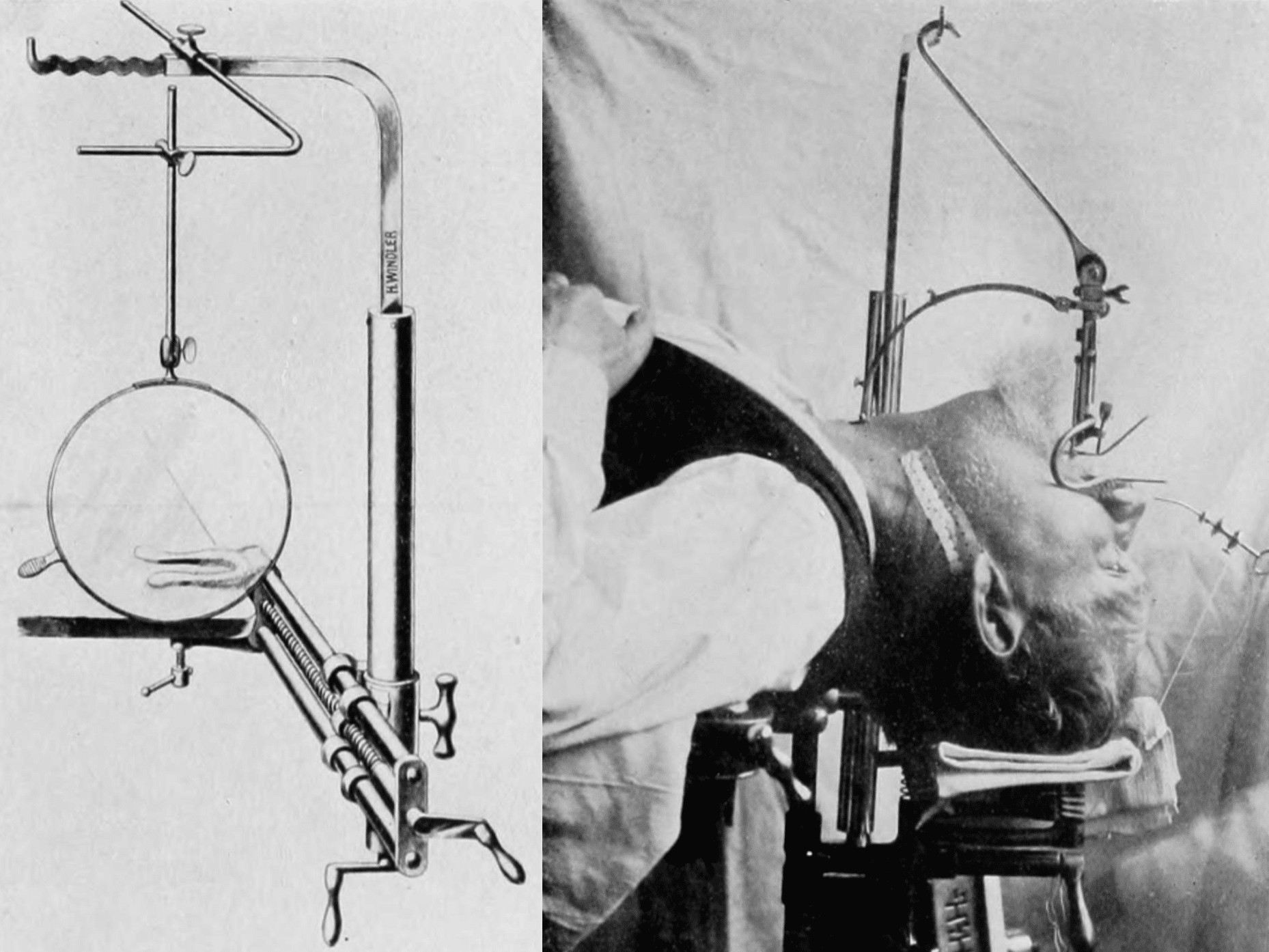
1915 – American laryngologist Robert Clyde Lynch (1880-1931) published Suspension laryngoscopy and its accomplishments. Lynch adopted Killian’s apparatus after seeing it demonstrated in 1913, but found its exposure of the anterior commissure inadequate in many adults. He strengthened and modified the gallows, mouth gag, tooth plates and spatulae, and altered the suspension geometry while retaining Killian’s underlying principle.
Lynch’s most important modification was to tilt the distal spatula anteriorly rather than maintain Killian’s relatively straight axis. This elevated the epiglottis and improved exposure of the anterior commissure, particularly in muscular or anatomically difficult patients.
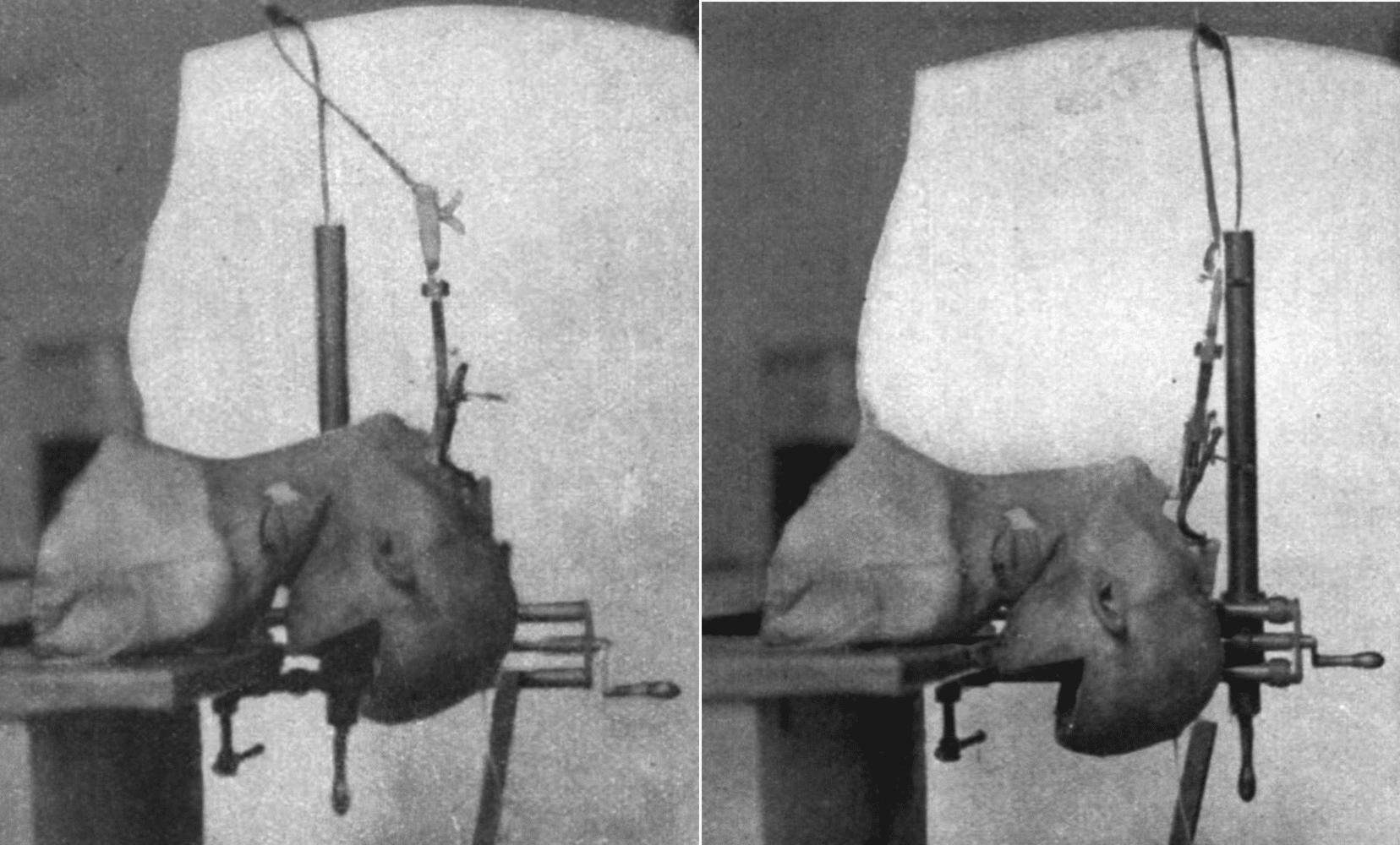
1916–1920 – Lynch continued to report his experience with suspension laryngoscopy and developed its use for precise bimanual dissection of intrinsic laryngeal tumours.
Eponymythology
Killian introduced the technique as Schwebelaryngoskopie (suspension laryngoscopy) and developed the original suspension gallows and instrumentation.
Lynch modified Killian’s apparatus and extended its operative use in the United States. The terms Lynch suspension laryngoscope and Killian–Lynch suspension laryngoscope were used for the equipment
Major Publications
- Killian G. Zur Anatomie der Parovarialcysten. Dissertation, 1895
- Killian G. Über die Bursa und Tonsilla pharyngea. 1888
- Killian G. Die Untersuchung der hinteren Larynxwand. 1890
- Killian G. Ueber directe Bronchoscopie. Münchener medizinische Wochenschrift, 1898, 45: 844-847. [Introduction of direct bronchoscopy]
- Killian G. Zur Geschichte der Oesophago- und Gastroskopie. Deutsche Zeitschrift für Chirurgie, 1900-1901, 58: 499-512. [Describes first use of oesophagoscope by Kussmaul]
- Killian G. Die submucöse Fensterresektion der Nasenscheidewand. Archiv für Laryngologie und Rhinologie, 1904; 16: 362-387
- Killian G. The mouth of the esophagus. Laryngoscope 1907; 17(6): 421–428
- Killian G. Die Schwebelaryngoscopie. Archiv für Laryngologie und Rhinologie, 1912, 26: 277-317.
- Killian G. Suspension laryngoscopy and its practical use, Semon lecture. 1914
- Killian G. Suspension laryngoscopy. In: Jackson C, Peroral endoscopy and laryngeal surgery. 1915: 133-154.
- Voss O, Killian G. Gehörorgan Obere Luft- und Speisewege. 1921
References
Biography
- Festschrift Gustav Killian am 2. Juni 1920 zu seinem 60. Geburtstage / von seinen Schüler und Freunden gewidmet. Archiv für Laryngologie und Rhinologie 1920; 33
- Killian H. Gustav Killian, sein Leben, sein Werk : Zugleich ein Beitrag zur Geschichte der Bronchologie und Laryngologie. 1958
- Zöllner F. Gustav Killian, father of bronchoscopy. Arch Otolaryngol. 1965 Dec;82(6):656-9.
- Dietzel K. 70 Jahre Bronchoskopie–in memoriam Gustav Killian. Z Erkr Atmungsorgane Folia Bronchol. 1969;130(1):61-4.
- Becker H. Gustav Killian: A Biographical Sketch. Journal of Bronchology. 1995;2(1):77-83.
- Plinkert PK. Gustav Killian–ein Pionier der Endoskopie. HNO. 1998 Jul;46(7):629-30.
- Prowse SJ, Makura Z. Gustav Killian: beyond his dehiscence. J Laryngol Otol. 2012 Nov;126(11):1164-8.
Eponymous terms
- Kollofrath O. Entfernung eines Knochenstücks aus dem rechten Bronchus auf natürlichem Wege und unter Anwendung der directen Laryngoskopie. Münchener medicinische Wochenschrift. 1897;44(38):1038–1039.
- Krauss L. Die Killian’sche Radicaloperation chronischer Stirnhöhleneiterungen. Archiv für Laryngologie und Rhinologie, Berlin, 1903; 13: 28-88.
- Lynch RC. Suspension laryngoscopy and its accomplishments. Ann Otol Rhinol laryngoI 1915; 24: 429-446.
- Lynch RC. A resume of my year’s work with suspension laryngoscopy. Trans Am Laryngol Assoc 1916; 38: 158-75.
- Lynch RC. Intrinsic carcinoma of the larynx, with a second report of the cases operated on by suspension and dissection. Trans Am Laryngol Assoc 1920; 40: 119-26.
- Breisinger A, Laszig R, Matthys H. Gustav Killian Gedächtnislesung. 100 Jahre Bronchoskopie–Vorgeschichte und Weiterentwicklung. Pneumologie. 1997 Jul;51(7):611-9.
- Zeitels S, Burns J, Dailey S. Suspension Laryngoscopy Revisited Annals of Otology, Rhinology & Laryngology. 2004;113(1):16-22.
- Hansson N, Halling T, Fangerau H. The Nobel Prize and otolaryngology: ‘Papa Gunnar’s’ promotion of his peers Gustav Killian and Themistocles Gluck’, Acta Oto-Laryngologica, 2016; 136(9): 871–874.
Eponym
the person behind the name

MB ChB, Edinburgh Medical school. F1/F2 in Bath, now in Australia, Perth preparing for a career in Emergency medicine. I have an interest in wilderness medicine and women’s health, but haven’t fully found my niche interest yet.

BA MA (Oxon) MBChB (Edin) FACEM FFSEM. Emergency physician, Sir Charles Gairdner Hospital. Passion for rugby, medical history, medical education, and asynchronous learning #FOAMed evangelist. Editor-in-Chief and Webmaster of Life in the Fast lane, LITFL | On Call 4e| Eponyms | Books | Horology |