


Hampton hump
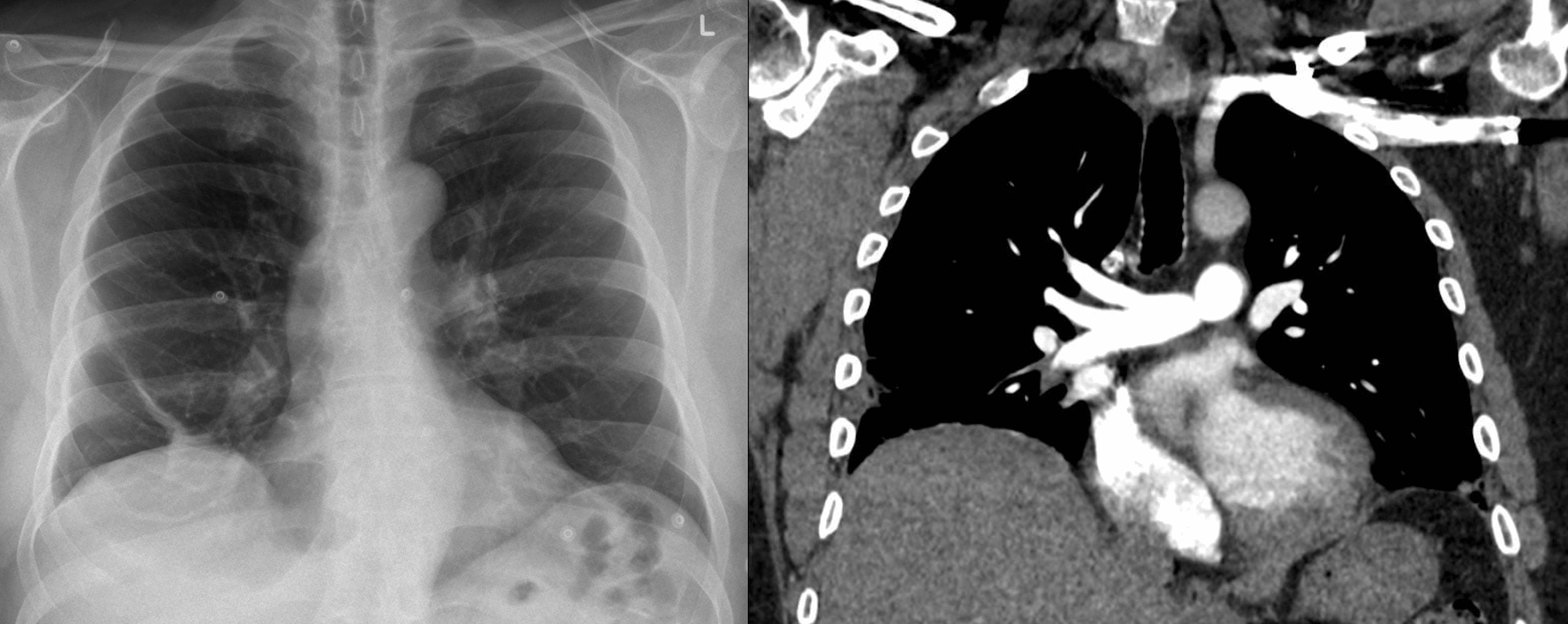
Hampton hump is a peripheral, pleural-based pulmonary opacity produced by haemorrhagic pulmonary infarction secondary to pulmonary embolism. It is classically described as wedge-shaped. However the infarct is a pleural-based rounded, hemispherical or truncated-cone consolidation whose inward margin may form a characteristic convex “hump” towards the hilum or heart.
Hampton hump most often occurs in the lower lung fields and may be associated with pleural effusion. It is an uncommon finding with low sensitivity for acute PE. When recognised in the appropriate clinical setting it should increase suspicion for pulmonary infarction and prompt definitive investigation rather than establish the diagnosis.
Hampton hump is not fundamentally “a wedge”. It is the radiographic expression of a pleural-based haemorrhagic infarct, with the characteristic hump derived from its convex inward margin.
Historical development
1940 – Aubrey Otis Hampton and Benjamin Castleman published Correlation of postmortem chest teleroentgenograms with autopsy findings with special reference to pulmonary embolism and infarction. Their study correlated postmortem radiographs with detailed gross and microscopic lung pathology and provided one of the foundational descriptions of the evolution and radiographic morphology of pulmonary infarction.
The series arose from more than 3,500 autopsies. They analysed 370 cases of pulmonary embolism and infarction, divided between postoperative, cardiac and non-cardiac medical patients. The frequency of infarction varied substantially with 58% of postoperative cases, 90% of cardiac cases and 62% of non-cardiac medical cases.
Hampton and Castleman demonstrated that during the first one to two days the affected lung showed marked capillary congestion and haemorrhage into the alveoli without established alveolar-wall necrosis. The corresponding radiographic opacity often did not become visible until at least 12 hours, and sometimes 24 hours, after symptom onset. Necrosis of the alveolar walls became evident after approximately the second day, followed by organisation and eventual contraction or scarring.
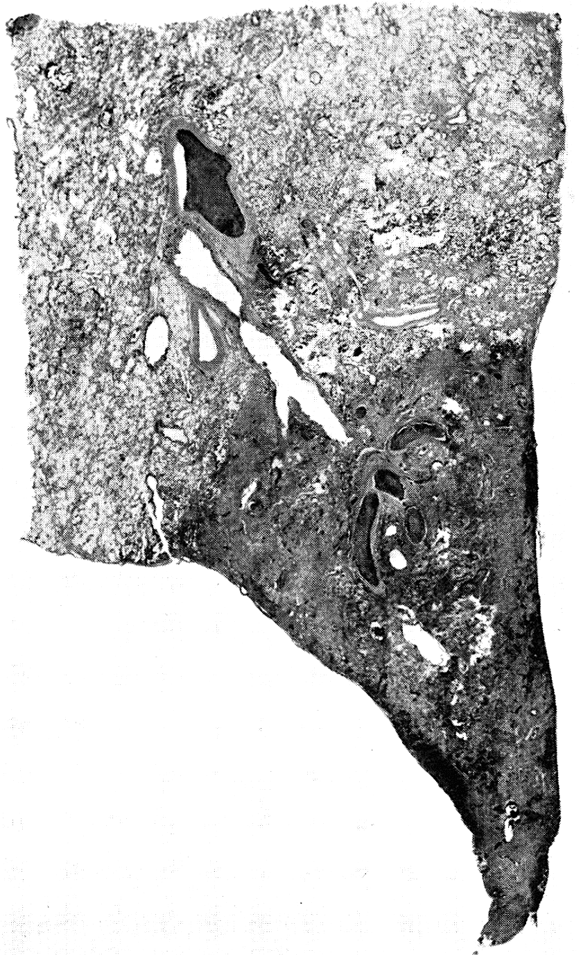
They described infarcts as peripheral and in contact with a pleural surface, with their long axis parallel to the involved pleura. Depending on anatomy and projection they might appear rounded, irregular, triangular or half-spindle shaped. The characteristic convex medial margin produced the hump-shaped shadow appearance.
The medial or cardiac margin of an infarct is convex toward the heart, presents a serrated margin, and, if the central roentgen ray is tangent to it, a a convex, slightly irregular “hump”-shaped shadow is produced. When the lateral costophrenic margins of the lung are involved, the posteroanterior view of the chest shows the “hump”-shaped shadow. When the anterior and posterior costophrenic margins are involved, the “hump” shadow is seen only in the lateral view.
Hampton 1940
1958 – Felix George Fleischner published Pulmonary embolism. He used the term “Hampton’s hump” when describing the convex contour of a pulmonary infarct, demonstrating that the eponym had entered radiological usage by this time.
1993 – Worsley et al., analysing the PIOPED cohort, prospectively evaluated the classic radiographic findings of PE. Hampton hump and pleural-based opacity proved insensitive, with later reviews reporting approximately 22% sensitivity and 82% specificity.
An absent Hampton hump provides no useful exclusion of PE. Its presence is suggestive of pulmonary infarction but remains non-diagnostic.
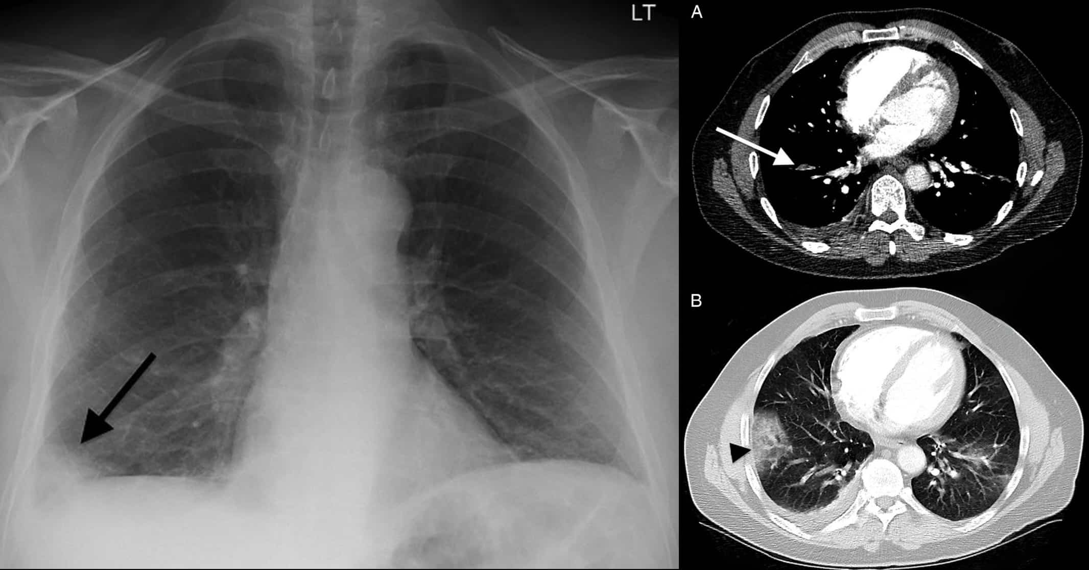
2014 – Patel et al. published Radiographic features of pulmonary embolism: Hampton’s hump. They demonstrated a peripheral wedge-shaped right lower-lobe opacity on chest radiography. CTPA showed an embolic filling defect in the supplying lateral basal pulmonary artery and a more peripheral wedge-shaped consolidation corresponding to the radiographic Hampton hump.
Eponymythology
Hampton and Castleman did not describe pulmonary infarction simply as a triangular or wedge-shaped opacity. They described infarcts as pleural-based lesions whose shape depends on the anatomy of the involved lung and the radiographic projection. They considered infarcts “rarely if ever actually triangular”. The characteristic feature giving rise to the later eponym was the convex medial or cardiac margin, which could produce a rounded “hump-shaped” shadow when viewed tangentially.
The original paper used descriptive language rather than the term Hampton’s hump. By 1958, however, Felix Fleischner was using “Hampton’s hump” for this radiographic appearance. Modern descriptions commonly reduce the sign to a “wedge-shaped pleural-based opacity”, which is clinically useful shorthand but loses some of the anatomical precision of Hampton and Castleman’s original description.
Associated Persons
- Nils Westermark (1892-1980) [Westermark sign]
- Aubrey Otis Hampton (1900-1955) [Hampton hump]
- Benjamin Castleman (1906–1982)
- Felix George Fleischner (1893-1969) [Fleischner sign and Fleischner lines]
- C. H. Joseph Chang (1929-2017) [Chang sign]
- Antonio Palla [Palla sign]
Alternative names
- Hampton’s hump
References
Original articles
- Hampton AO, Castleman B. Correlation of postmortem chest teleroentgenograms with autopsy findings with special reference to pulmonary embolism and infarction. American journal of roentgenology and radium therapy. 1940; 43: 305 –326. [Hampton Hump]
Eponym review
- Worsley DF, Alavi A, Aronchick JM, Chen JT, Greenspan RH, Ravin CE. Chest radiographic findings in patients with acute pulmonary embolism: observations from the PIOPED Study. Radiology. 1993 Oct;189(1):133-6.
- Piazza G, Goldhaber SZ. Acute pulmonary embolism: part I: epidemiology and diagnosis. Circulation. 2006 Jul 11;114(2):e28-32.
- Marshall GB, Farnquist BA, MacGregor JH, Burrowes PW. Signs in thoracic imaging. J Thorac Imaging. 2006 Mar;21(1):76-90.
- Stein PD, et al; PIOPED II Investigators. Diagnostic pathways in acute pulmonary embolism: recommendations of the PIOPED II Investigators. Radiology. 2007 Jan;242(1):15-21.
- Pipavath SN, Godwin JD. Acute pulmonary thromboembolism: a historical perspective. AJR Am J Roentgenol. 2008 Sep;191(3):639-41.
- Taylor BT, Pezzo SP, Rumbak M. Palla’s sign and Hampton’s hump in pulmonary embolism. Respiration. 2010;80(6):568.
- Ladeiras-Lopes R, Neto A, Costa C, Sousa M, Ferreira P, Dias VP, Ribeiro VG. Hampton’s hump and Palla’s sign in pulmonary embolism. Circulation. 2013 May 7;127(18):1914-5
- Patel UB, Ward TJ, Kadoch MA, Cham MD. Radiographic features of pulmonary embolism: Hampton’s hump. Postgraduate Medical Journal, 2014; 90(1065): 420–421.
- Miniati M, Bottai M, Ciccotosto C, Roberto L, Monti S. Predictors of Pulmonary Infarction. Medicine (Baltimore). 2015 Oct;94(41):e1488.
- Hsu CW, Su HY. Palla’s sign and Hampton’s hump in pulmonary embolism. QJM. 2017 Jan;110(1):49-50.
- Cadogan M. CXR eponyms in pulmonary embolism. LITFL
eponymictionary
the names behind the name

BA MA (Oxon) MBChB (Edin) FACEM FFSEM. Emergency physician, Sir Charles Gairdner Hospital. Passion for rugby, medical history, medical education, and asynchronous learning #FOAMed evangelist. Editor-in-Chief and Webmaster of Life in the Fast lane, LITFL | On Call 4e| Eponyms | Books | Horology |