


Fleischner lines
Fleischner lines are linear or band-like pulmonary opacities produced by focal plate-like (discoid) atelectasis viewed edge-on on chest radiography. They are most often seen in the lower lung fields, where they may be horizontally orientated, but their direction depends on the orientation of the affected pulmonary subsegment.
Classically, Fleischner lines are several centimetres long and a few millimetres thick, extend to a pleural surface, and do not cross an interlobar fissure. They represent atelectatic lung rather than an occluded pulmonary artery or pulmonary infarct itself. Baron described typical lines as approximately 2–7 cm long and 2–7 mm thick.
Fleischner lines are non-specific. They may occur with mucus plugging, aspiration, pneumonia, abdominal disease, peripheral tumour and other causes of focal hypoventilation or bronchial obstruction.
In pulmonary embolism they are an indirect radiographic manifestation, historically attributed to obstruction of small bronchi by retained secretions or haemorrhagic exudate with subsequent focal atelectasis. They are now regarded as an uncommon radiographic clue rather than a diagnostic sign of PE. The term is distinct from the Fleischner sign, which describes enlargement of a proximal pulmonary artery associated with pulmonary embolism.
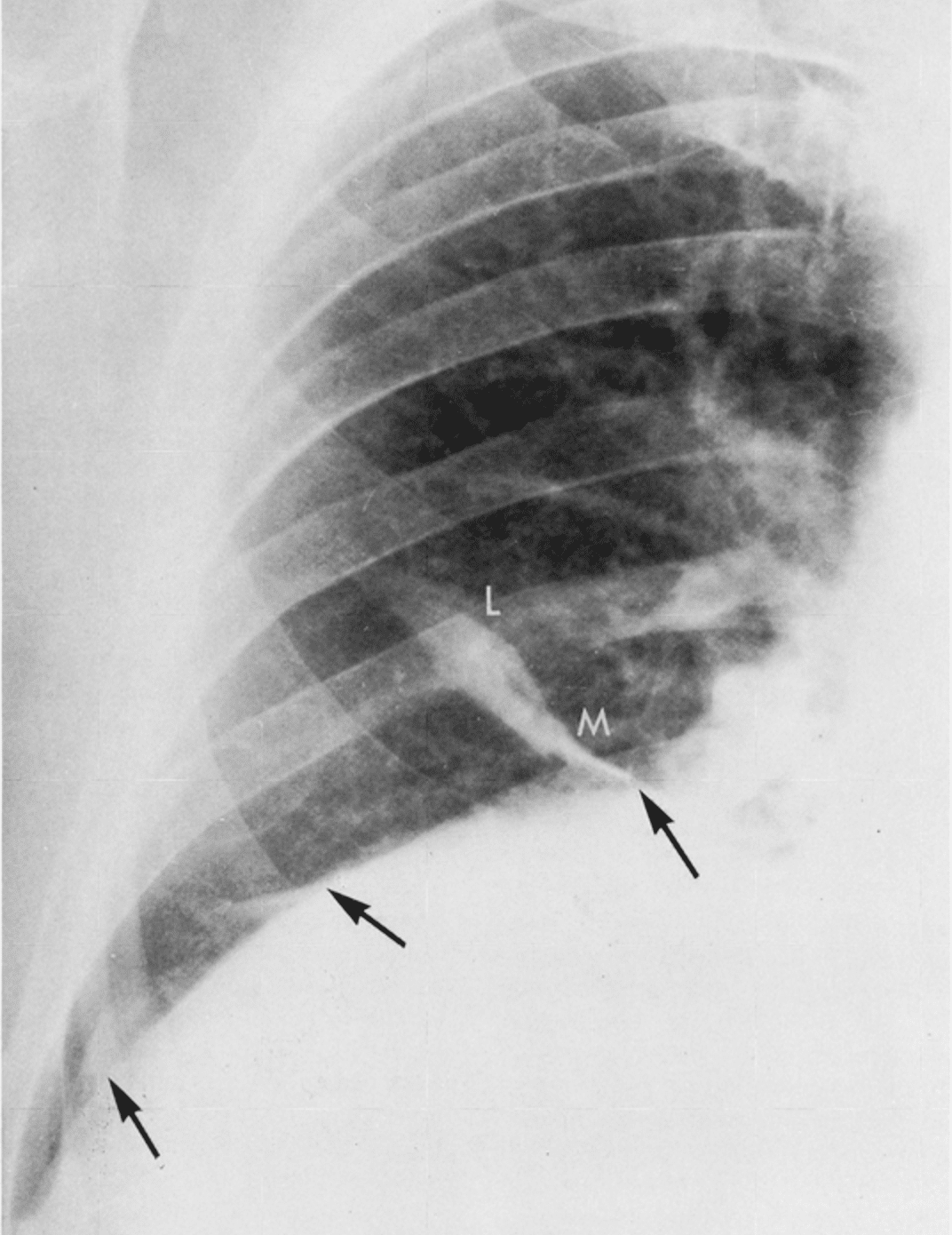
History of the Fleischner line
1936 – Felix George Fleischner published three papers investigating pulmonary collapse, plate-like atelectasis and basal horizontal pulmonary shadows. These establish the basis of the radiographic findings later termed Fleischner lines.
1941 – Fleischner, Aubrey Otis Hampton and Benjamin Castleman published Linear shadows in the lung (interlobar pleuritis, atelectasis and healed infarction). They distinguished plate-like atelectasis from linear shadows caused by pleural disease and healing/healed pulmonary infarction.
1958-1962 – Fleischner incorporated plate atelectasis into his broader description of impaired ventilation accompanying pulmonary embolism, associating elevated diaphragm, reduced diaphragmatic excursion and basal plate atelectasis.
1972 – Murray G. Baron published Fleischner lines and pulmonary emboli stating that the linear shadows were “commonly referred to as Fleischner lines”. He defined their morphology and emphasised that they were an indirect, non-specific manifestation of PE caused by focal bronchial obstruction and atelectasis rather than by direct visualisation of vascular occlusion.
1982 – Donald G. Shaw published Fleischner’s lines-a model? noting continuing disagreement over bronchial versus vascular origins. He proposed that compression of relatively non-compliant lung by the diaphragm could itself produce basal plate-like atelectatic shadows.
2019 – Zuin et al. reported Fleischner lines in acute PE with distal pulmonary infarction, describing them as rare and non-specific long bands of focal atelectasis, typically basal and horizontally orientated. Their CXR demonstrates the lines with CT confirmation of segmental PE and infarction.
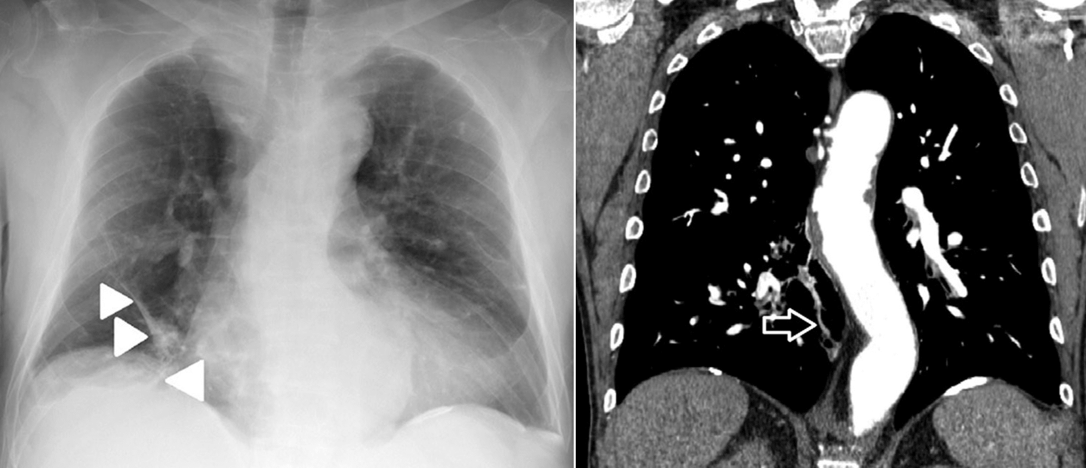
2023 – Yudin retained discoid/linear/plate/band atelectasis and Fleischner lines as synonymous modern radiological terminology, emphasising their plate-like morphology and broad differential diagnosis.
Associated Persons
- Nils Westermark (1892-1980) [Westermark sign]
- Aubrey Otis Hampton (1900-1955) [Hampton hump]
- Benjamin Castleman (1906–1982)
- Felix George Fleischner (1893-1969) [Fleischner sign and Fleischner lines]
- C. H. Joseph Chang (1929-2017) [Chang sign]
- Antonio Palla [Palla sign]
Alternative names
- Fleischner lines, Fleischner’s lines
- Plate-like atelectasis, discoid atelectasis, linear atelectasis
References
Original articles
- Fleischner F. Atelektase und gerichteter Kollaps der Lunge. Fortschritte auf dem Gebiete der Röntgenstrahlen und der Nuklearmedizin, 1936, 53, 607-625.
- Fleischner F. Plattenformige Atelektasen in der Unterlappen der Lunge. Fortschritte auf dem Gebiete der Röntgenstrahlen und der Nuklearmedizin 1936; 54: 315-321.
- Fleischner F. Über das Wesen der basalen horizontalen Schattenstreifen im Lungenfeld. Wiener Archiv für innere Medizin 1936; 28: 461-480.
- Fleischner F, Hampton AO, Castleman B. Linear shadows in the lung (interlobar pleuritis, atelectasis and healed infarction). Am J Roentgenol 1941; 46(5): 610-618
- Fleischner FG. Pulmonary embolism. Canadian Medical Association Journal. 1958; 78(9): 653–660.
- Fleischner FG. Roentgen diagnosis of pulmonary embolism. Heart Bull. 1961 Nov-Dec;10:104-7.
- Fleischner FG. Pulmonary embolism. Clin Radiol. 1962 Jul;13:169-82
Eponym review
- Williams JR, Wilcox WC. Pulmonary embolism: roentgenographic and angiographic considerations. AJR Am J Roentgenol. 1963; 89: 333.
- Shaw DG. Fleischner’s lines-a model? Br J Dis Chest. 1982 Jan;76(1):97-8.
- Baron MG. Fleischner lines and pulmonary emboli. Circulation. 1972 Jan;45(1):171-8.
- Worsley DF et al. Chest Radiographic Findings in Patients With Acute Pulmonary Embolism: Observations From the PIOPED Study. Radiology. 1993 Oct;189(1):133-6.
- Elliott CG, Goldhaber SZ, Visani L, DeRosa M. Chest radiographs in acute pulmonary embolism. Results from the International Cooperative Pulmonary Embolism Registry. Chest. 2000; 118(1): 33-8.
- Marshall GB, Farnquist BA, MacGregor JH, Burrowes PW. Signs in thoracic imaging. J Thorac Imaging. 2006; 21(1): 76-90.
- Zuin M, Rigatelli G, Zonzin P, Roncon L. Chest X-ray in acute pulmonary embolism: forgiven but not forgotten. Intensive Care Med. 2019 Dec;45(12):1822-1823.
- Yudin A. Discoid (Linear, Plate, or Band) Atelectasis, Fleischner Lines. In: Metaphorical Signs in Computed Tomography of Chest and Abdomen. Springer 2023
- Cadogan M. CXR eponyms in pulmonary embolism. LITFL
eponymictionary
the names behind the name

BA MA (Oxon) MBChB (Edin) FACEM FFSEM. Emergency physician, Sir Charles Gairdner Hospital. Passion for rugby, medical history, medical education, and asynchronous learning #FOAMed evangelist. Editor-in-Chief and Webmaster of Life in the Fast lane, LITFL | On Call 4e| Eponyms | Books | Horology |