![]()
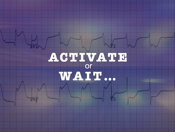
Activate or Wait – 002
Further reading

![]()
Further reading
8 deadly ECG patterns NOT to miss -- Part 2, the occlusion version. These patients require immediate cardiology referral for emergent reperfusion therapy.
There are five features on this "normal" ECG that suggest impending inferior STEMI - can you spot them?
Can ST depression and T wave inversion in aVL be normal? Can BER cause reciprocal changes? Learn about using the QRS-T wave angle to answer these questions
With worked examples in the next three posts, we look at ways to recognise early ECG features of OMI before waiting for a "STEMI" to evolve
With a great case example, we discuss diagnosing OMI in the presence of intraventricular conduction delay and/or prior anterior myocardial infarction
This characteristic ECG pattern should be in every critical care practitioner's knowledge base as a STEMI-equivalent, regardless of the magnitude of ST-segment changes seen
Aslanger et al identified a specific ECG pattern concerning for acute inferior occlusion MI in patients with concomitant multi-vessel disease, that does not display contiguous ST-segment elevation or fulfil STEMI criteria
The OMI/NOMI paradigm: Better recognising patients with acute ischaemia that will respond to Percutaneous Coronary Intervention (PCI)