
![]()
ECG Case 064
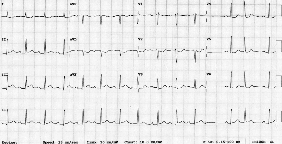
Chest pain and diaphoresis. BP 80/50. Describe and interpret his ECG
Describe and interpret this ECG
ECG ANSWER and INTERPRETATION
Key Abnormalities
- There is ST elevation in the inferior leads II, III and aVF.
- The concave morphology might lead you to suspect pericarditis – however, there is reciprocal change in the high lateral leads I and aVL, confirming the diagnosis of inferior STEMI.
There are additional features suggestive of right ventricular infarction:
- ST elevation in III > II
- Isoelectric ST segment in V1 with ST depression in V2
Other Abnormalities
- There is a break in the rhythm towards the end of the rhythm strip, with what appears to be a non-conducted P wave, suggesting the development of 2nd degree AV block — e.g. a slowly-evolving Wenckbach cycle.
- The 13th QRS complex appears to be a supraventricular ectopic beat (PAC or PJC).
References
Further Reading
- Wiesbauer F, Kühn P. ECG Mastery: Yellow Belt online course. Understand ECG basics. Medmastery
- Wiesbauer F, Kühn P. ECG Mastery: Blue Belt online course: Become an ECG expert. Medmastery
- Kühn P, Houghton A. ECG Mastery: Black Belt Workshop. Advanced ECG interpretation. Medmastery
- Rawshani A. Clinical ECG Interpretation ECG Waves
- Smith SW. Dr Smith’s ECG blog.
- Wiesbauer F. Little Black Book of ECG Secrets. Medmastery PDF
TOP 100 ECG Series
Emergency Physician in Prehospital and Retrieval Medicine in Sydney, Australia. He has a passion for ECG interpretation and medical education | ECG Library |