![]()
Abdominal CT: Cystogram
Ordering a cystogram
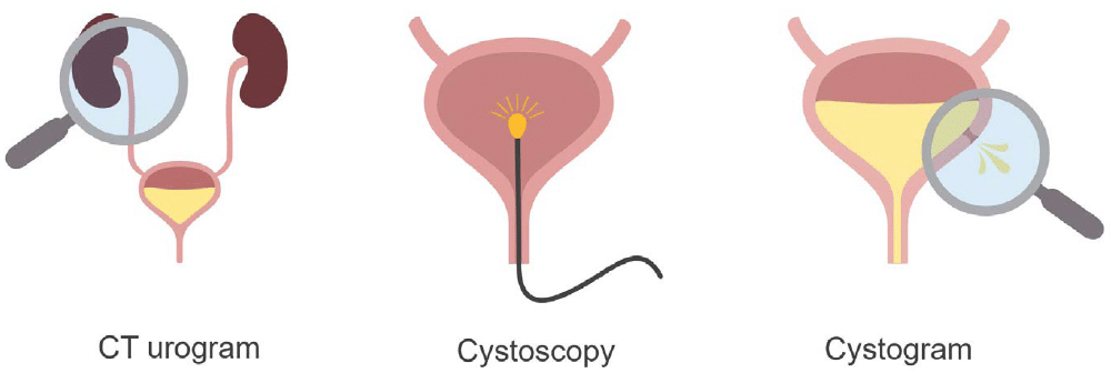
While a CT urogram is a great way to visualize the upper genitourinary tract, when you want to evaluate just the bladder using imaging, a CT cystogram is your best option.
When a patient has blood in their urine, they are routinely evaluated using two exams:
- CT urogram: This exam evaluates the upper genitourinary tract.
- Optical cystoscopy: Performed by a urologist to examine the inner lining of the bladder for a subtle tumour difficult to visualise on CT
Like a cystoscopy, a CT cystogram is also designed to look at the bladder, but it is best for evaluating leaks related to surgery or injury
How is a cystogram performed?
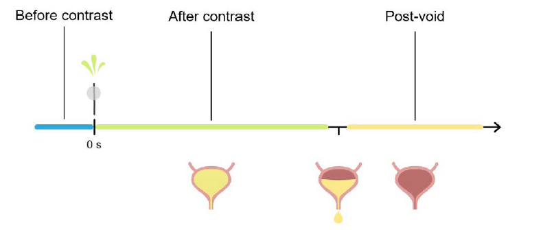
The cystogram does not use IV contrast. Instead, a urinary catheter is inserted and contrast is infused into the bladder using gravity. This continues until there is a sensation of fullness and slight discomfort, which indicates contraction of the bladder muscle. It is important to reach this point because some bladder injuries do not become visible before this contraction.
Images can be obtained at three time points:
- Before contrast
- After contrast fills the bladder
- After emptying the bladder (post-void imaging)
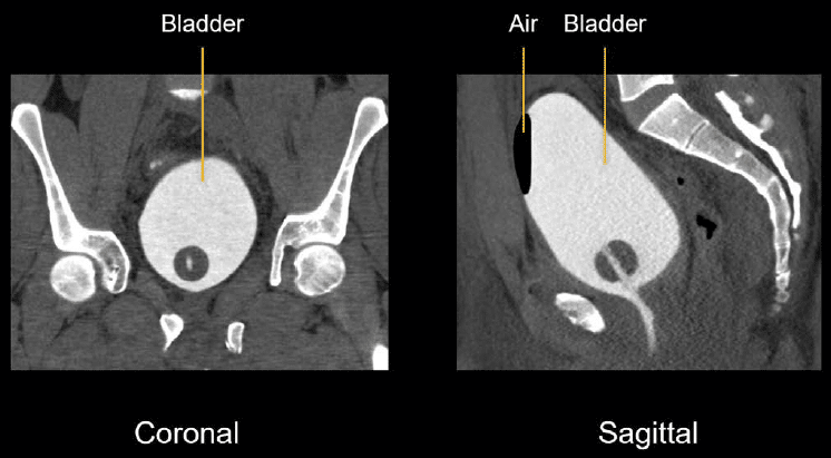
Example 1: Normal cystogram
Reviewing cystogram images When reviewing cystogram images, you will expect to see a smooth bladder wall without any contrast leaks.
Example 2: intraperitoneal bladder rupture
The images below are taken from a trauma patient involved in a high velocity motor vehicle collision and came to the hospital with pelvic fractures and a possible bladder injury.
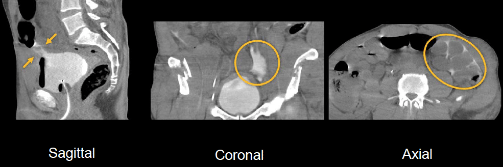
- Sagittal image: Foley catheter filling the bladder with dense contrast. There is a sizeable defect near the top of the bladder where contrast can be seen leaking out.
- Coronal view: again, we can see contrast leaking out of the bladder.
- Axial image: you can see contrast surrounding the bowel loops in the abdominal cavity
This injury is an intraperitoneal bladder rupture and requires surgical repair.
Example 3: fistulae
A cystogram can also be used to look for abnormal connections between two structures. When this occurs between the bladder and rectum, it’s called a colovesical fistula. When it occurs between the bladder and vagina, it’s called a vesicovaginal fistula.
A fistula can form as a result of chronic inflammation, surgery, or radiation therapy. A cystogram can help to determine the size and configuration of the fistula and be used for planning surgical repair.
The following patient had a history of rectal cancer and radiation therapy and presented with urine leaking from the rectum, suggesting a fistula.
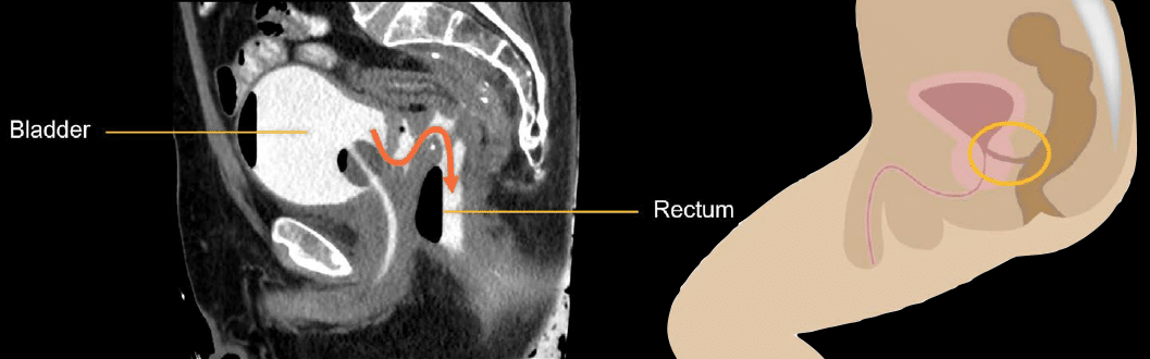
As you can see in this cystogram image, the fistula appears thickened, forming an irregular tract between the bladder and rectum.
This is an edited excerpt from the Medmastery course Abdominal CT Essentials by Michael P. Hartung, MD. Acknowledgement and attribution to Medmastery for providing course transcripts.
- Hartung MP. Abdominal CT: Common Pathologies. Medmastery
- Hartung MP. Abdominal CT: Essentials. Medmastery
- Hartung MP. Abdomen CT: Trauma. Medmastery
References
- Top 100 CT scan quiz. LITFL
Radiology Library: Abdominal CT Basics
- Hartung MP. Abdominal CT: Phases. LITFL
- Hartung MP. Abdominal CT: Cancer staging. LITFL
- Hartung MP. Abdominal CT: GU imaging. LITFL
- Hartung MP. Abdominal CT: Urogram. LITFL
- Hartung MP. Abdominal CT: Cystogram. LITFL
- Hartung MP. Abdominal CT: Flank pain imaging. LITFL
Abdominal CT interpretation

Assistant Professor of Abdominal Imaging and Intervention at the University of Wisconsin Madison School of Medicine and Public Health. Interests include resident and medical student education, incorporating the latest technology for teaching radiology. I am also active as a volunteer teleradiologist for hospitals in Peru and Kenya. | Medmastery | Radiopaedia | Website | Twitter | LinkedIn | Scopus
BA MA (Oxon) MBChB (Edin) FACEM FFSEM. Emergency physician, Sir Charles Gairdner Hospital. Passion for rugby; medical history; medical education; and asynchronous learning #FOAMed evangelist. Co-founder and CTO of Life in the Fast lane | On Call: Principles and Protocol 4e| Eponyms | Books |