![]()
Abdominal CT: Urogram
Ordering a urogram
Let’s consider the scenario of an older patient presenting with blood in the urine. We not only have to think about the possibility of renal stones as the cause of the bleeding but also the possibility of renal tumours or urothelial tumours in the collecting system, ureters, and bladder.
In this case, the CT urogram is the exam of choice as it allows you to see the entire genitourinary tract by including three series:
- Non-contrast
- Nephrographic phase
- Excretory phase
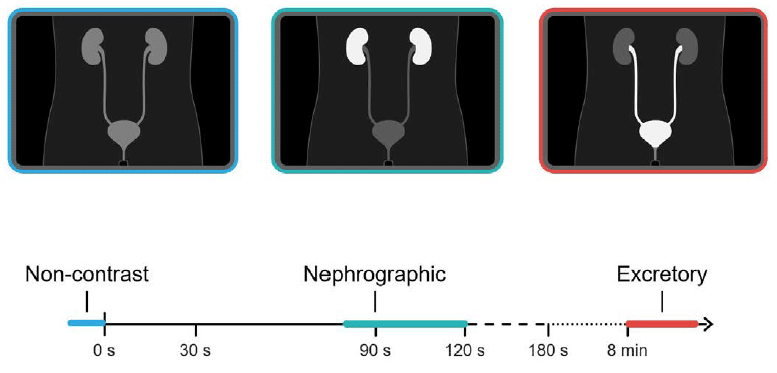
Nephrographic phase
The nephrographic phase is very similar to the portal venous phase, only slightly later at about 80–120 seconds after contrast is given. The phase provides the best view of the renal cortex to look for masses, but it is also great for looking at the other abdominal organs and bowel
Excretory phase
The excretory phase images are even more delayed, at eight or more minutes after contrast is given. This delay allows the contrast to mix with the urine excreted by the kidneys, making it bright, and enabling you to see the inside of the collecting system, ureters, and bladder in order to evaluate for masses.
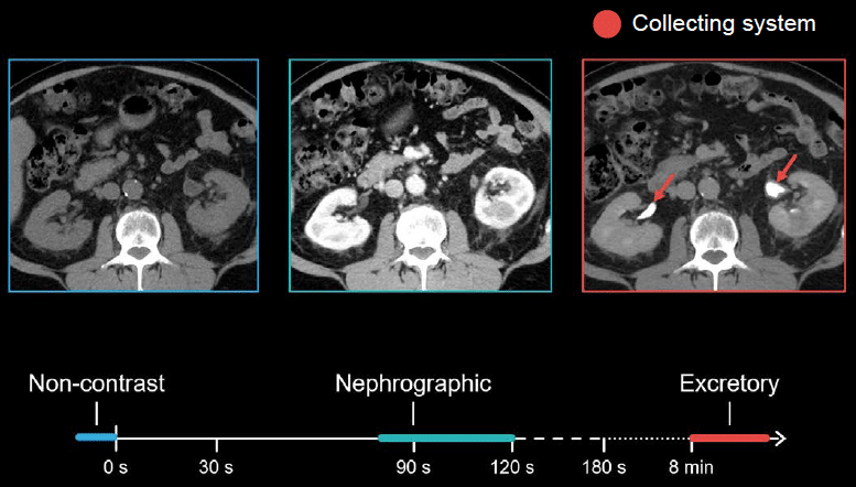
Example 1
- In the non-contrast image below, the kidneys look relatively gray and blend in with other soft tissue structures.
- In the nephrographic phase, the kidneys really light up and give us the best chance to evaluate the tissue for abnormal enhancement or masses.
- Finally, after waiting eight minutes, the collecting system has filled with contrast-opacified urine, which allows us to see the inside of the urinary system, from the ureters to the bladder.
Because a CT urogram requires multiple series, it is best to approach your review of this examination as a checklist, with each series providing essential information.
Checklist for kidney stones and kidney masses
- Kidney stones
- Kidney masses
- Collecting system
- Ureters
- Bladder
This is an edited excerpt from the Medmastery course Abdominal CT Essentials by Michael P. Hartung, MD. Acknowledgement and attribution to Medmastery for providing course transcripts.
- Hartung MP. Abdominal CT: Common Pathologies. Medmastery
- Hartung MP. Abdominal CT: Essentials. Medmastery
- Hartung MP. Abdomen CT: Trauma. Medmastery
References
- Top 100 CT scan quiz. LITFL
Radiology Library: Abdominal CT Basics
- Hartung MP. Abdominal CT: Phases. LITFL
- Hartung MP. Abdominal CT: Cancer staging. LITFL
- Hartung MP. Abdominal CT: GU imaging. LITFL
- Hartung MP. Abdominal CT: Urogram. LITFL
- Hartung MP. Abdominal CT: Cystogram. LITFL
- Hartung MP. Abdominal CT: Flank pain imaging. LITFL
Abdominal CT interpretation

Assistant Professor of Abdominal Imaging and Intervention at the University of Wisconsin Madison School of Medicine and Public Health. Interests include resident and medical student education, incorporating the latest technology for teaching radiology. I am also active as a volunteer teleradiologist for hospitals in Peru and Kenya. | Medmastery | Radiopaedia | Website | Twitter | LinkedIn | Scopus
BA MA (Oxon) MBChB (Edin) FACEM FFSEM. Emergency physician, Sir Charles Gairdner Hospital. Passion for rugby; medical history; medical education; and asynchronous learning #FOAMed evangelist. Co-founder and CTO of Life in the Fast lane | On Call: Principles and Protocol 4e| Eponyms | Books |