![]()
Abdominal CT: large intestine
Running the large bowel and appendix
Large bowel
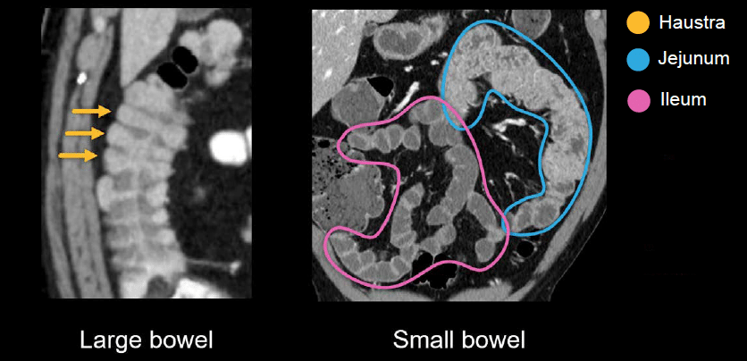
As you learn how to review the GI tract, you will become increasingly comfortable with the fold pattern of the large and small bowel. The distinguishing feature of the large bowel is haustra – incomplete folds that are spaced farther apart than the folds seen in the small bowel.
On CT, the appearance of the small bowel can range from the feathery folds of the jejunum in the left-upper and mid abdomen to the relatively smooth wall of the ileum in the lower abdomen.
Appendix
Trying to find the appendix on CT is an important goal of every scan. Review this set of PACS images in and try it for yourself.
There is a good chance you will be successful in finding the appendix by simply scrolling immediately to the right lower quadrant and looking for a thin, tubular structure (or a dilated, angry-looking one if the patient has acute appendicitis).
However, this doesn’t leave you with many problem-solving options if you can’t find the appendix. So, let’s consider a more dependable approach.
Begin at the end
The most reliable way of running the lower GI tract is to begin at the end (i.e., at the anal canal) and work your way backward to the cecum. On your way, you should watch for the following, as these can indicate infection, inflammation, ischemia, or a mass:
- Diverticular disease
- Abnormally increased or decreased enhancement
- Wall thickening
Using the same PACS images
- Take a look at the axial images and find the anal canal. For there, we can run the large bowel in reverse.
- Scrolling up from the anal canal, we run through the rectum, the s-shaped sigmoid colon, and the descending colon.
- In the left upper quadrant, we find the splenic flexure, then across the midline we can follow the transverse colon.
- In the right upper quadrant, we find the hepatic flexure, and for the home stretch we can follow the ascending colon down to the cecum.
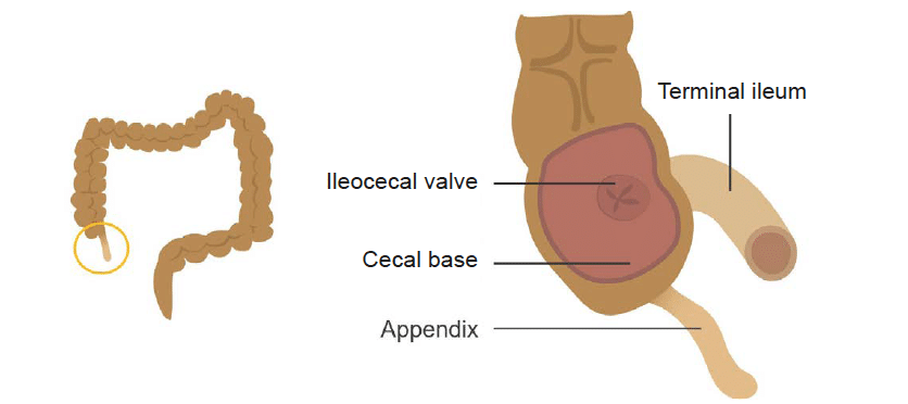
- Finally, we can see the appendix arising from the cecal base.
Important landmarks
There are key landmarks in the right lower quadrant that help us to locate structures of interest. For example, when looking for the appendix, first find the terminal ileum and follow it into the ileocaecal valve. From there, go down a few cm to the caecal base, which is where you can expect to find the appendix
Let’s get some practice identifying these important right lower quadrant landmarks. Review these PACS images and:
- Take a look at the coronal images and find the distal and terminal ileum.
- From there, you can follow into the fatty rind of the ileocaecal valve and into the cecum.
- Going down a few cm, you can find the cecal base, and then scroll back and forth to find the appendix.
This is an edited excerpt from the Medmastery course Abdominal CT Essentials by Michael P. Hartung, MD. Acknowledgement and attribution to Medmastery for providing course transcripts.
- Hartung MP. Abdominal CT: Common Pathologies. Medmastery
- Hartung MP. Abdominal CT: Essentials. Medmastery
- Hartung MP. Abdomen CT: Trauma. Medmastery
References
- Top 100 CT scan quiz. LITFL
Radiology Library: Abdominal CT: Imaging the abdominal organs and bowel
- Hartung MP. Abdominal CT: Evaluating the liver
- Hartung MP. Abdominal CT: Evaluating the biliary system and pancreas
- Hartung MP. Abdominal CT: Imaging the spleen and adrenal glands
- Hartung MP. Abdominal CT: Mastering the genitourinary system
- Hartung MP. Abdominal CT: Evaluating the lower oesophagus and stomach
- Hartung MP. Abdominal CT: Imaging the small bowel
- Hartung MP. Abdominal CT: Viewing the peritoneal cavity
- Hartung MP. Abdominal CT: Running the large bowel and appendix
Abdominal CT interpretation

Assistant Professor of Abdominal Imaging and Intervention at the University of Wisconsin Madison School of Medicine and Public Health. Interests include resident and medical student education, incorporating the latest technology for teaching radiology. I am also active as a volunteer teleradiologist for hospitals in Peru and Kenya. | Medmastery | Radiopaedia | Website | Twitter | LinkedIn | Scopus

MBChB (hons), BMedSci - University of Edinburgh. Living the good life in emergency medicine down under. Interested in medical imaging and physiology. Love hiking, cycling and the great outdoors.