Activate or Wait – 009
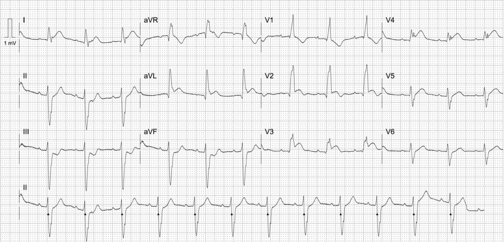
81-year-old female with three hours of central, crushing chest pain. Background of hypertension, heavy smoker.
We are 10 minutes from your tertiary centre.

Would you activate your cath lab/STEMI protocol?
ECG interpretation
Anteroseptal OMI
- Concordant ST elevation in leads V2-4, I and aVL — note the presence of right bundle branch block (RBBB)
- Concurrent left anterior fascicular block indicates a bifascicular block
- Reciprocal concordant ST depression in inferior leads II, III, aVF
- Evolving Q waves in aVR, aVL and V1-2
Although it may be pre-existing, a new bifascicular often accompanies a proximal LAD occlusion, as the right bundle branch and left anterior fascicle are supplied by a proximal septal branch of the LAD. These cases are EXTREMELY high risk – in our experience they are all post-arrest, pre-arrest, or in cardiogenic shock. They often have downsloping ST elevation that appears very atypical.
ST elevation in anterior leads here is prominent, however in the presence of a right bundle branch block, any degree of concordant ST elevation is highly concerning for occlusion myocardial infarction (OMI).
AI interpretation by PMcardio

PMcardio’s AI ECG model identified a STEMI/STEMI equivalent.
The explainability feature offers insights into problematic patterns in the respective leads, providing a clear rationale for the diagnosis.
Further reading
Outcome
Key Finding:
40% occlusion of proximal LAD – likely plaque rupture with thrombus but no occlusive disease.
Findings:
- Left Main Coronary Artery -Normal
- Left Anterior Descending Coronary Artery – 40% after LADD2
- Left Circumflex Coronary Artery – 30% proximal
- Right Coronary Artery -Dominant, irregular
- Left Ventriculogram – anterior hypokinesis with mild LV impairment
Plan:
- Likely plaque rupture LAD with thrombus but no occlusive disease
Clinical Pearls
Whilst there is no evidence-based equivalent of “Sgarbossa criteria” to diagnose acute coronary occlusion in right bundle branch block (RBBB), the same principles are applicable.
In RBBB with RSR’ in V1-3, we expect discordant ST depression. As such, any concordant ST elevation should be assumed to be OMI until proven otherwise. Examine closely as ST elevation is often downsloping and difficult to perceive.
New bifascicular block is highly associated with proximal LAD occlusion and negative outcomes. Raise suspicion for OMI and anticipate imminent clinical deterioration.
References
Further reading
- Burns E, Buttner R. Bifascicular Block. LITFL
Online resources
- Wiesbauer F, Kühn P. ECG Mastery: Yellow Belt online course. Understand ECG basics. Medmastery
- Wiesbauer F, Kühn P. ECG Mastery: Blue Belt online course: Become an ECG expert. Medmastery
- Kühn P, Houghton A. ECG Mastery: Black Belt Workshop. Advanced ECG interpretation. Medmastery
- Smith SW. Dr Smith’s ECG blog.
- Rawshani A. Clinical ECG Interpretation ECG Waves
ACTIVATE or WAIT
EKG Interpretation
MBBS DDU (Emergency) CCPU. Adult/Paediatric Emergency Medicine Advanced Trainee in Melbourne, Australia. Special interests in diagnostic and procedural ultrasound, medical education, and ECG interpretation. Co-creator of the LITFL ECG Library. Twitter: @rob_buttner
Dr. Stephen W. Smith is a faculty physician in the Emergency Medicine Residency at Hennepin County Medical Center (HCMC) in Minneapolis, MN, and Professor of Emergency Medicine at the University of Minnesota. Author of Dr Smith's ECG Blog | Bibliography | X |
Dr. Robert Herman is the Co-founder and Chief Medical Officer of PMcardio by Powerful Medical. He is a cardiovascular researcher at the Cardiovascular Center Aalst in Belgium, specializing in applying AI to enhance the detection and referral of cardiovascular patients. LinkedIn | X (formerly Twitter) | Get in Touch


