
![]()
Right Bundle Branch Block (RBBB)
ECG Diagnostic criteria
- QRS duration > 120ms
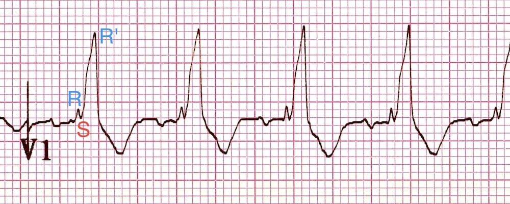
- RSR’ pattern in V1-3 (“M-shaped” QRS complex)
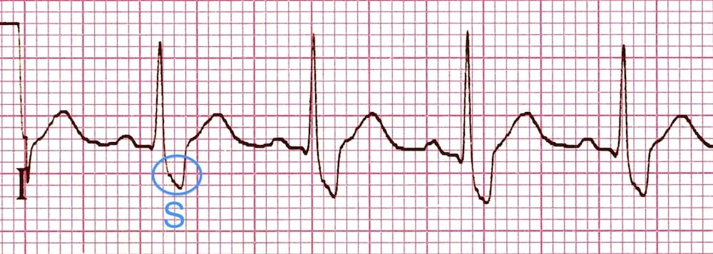
- Wide, slurred S wave in lateral leads (I, aVL, V5-6)

V1: RSR’ pattern in V1, with (appropriate) discordant T wave changes
V6: Widened, slurred S wave in V6
Associated features incude:
- Appropriate discordance with ST depression and/or T-wave inversion in right precordial leads (V1-3).
Electrophysiology
In normal cardiac conduction, impulses travel equally down the left and right bundles, with the septum activated from left to right and the formation of small Q waves in lateral leads
- In RBBB, the left ventricle is activated normally, thus the early part of the QRS complex correlating to septal depolarisation is unchanged
- There is delayed activation of the right ventricle as depolarisation originates from the left ventricle across the septum. This produces a secondary R wave (R’) in the precordial leads, and a wide, slurred S wave in lateral leads
- Normal activation of the left ventricle means that cardiac axis remains normal in isolated RBBB
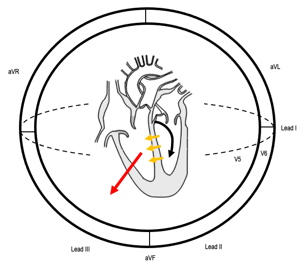
1) Left ventricular activation via the left bundle (black arrow) occurs normally
2) Septal depolarisation (yellow arrows) is thus unaffected, producing a normal early QRS complex
3) Activation of the RV originates across the septum. The resultant depolarisation vector (red arrow) produces delayed R waves in leads V1-3, and S waves in lateral leads
ECG QRS Morphology
QRS Morphology in V1
Sometimes rather than an RSR’ pattern in V1, there may be a broad monophasic R wave or a qR complex.
QRS Morphology in Lateral Leads
Appropriate discordance
- Appropriate discordance refers to the fact that abnormal depolarisation should be followed by abnormal repolarisation, which appears discordant to the preceding QRS complex
- In RBBB, this manifests as ST depression and/or T-wave inversion in leads V1-3
Causes of Right Bundle Branch Block
- Right ventricular hypertrophy / cor pulmonale
- Pulmonary embolus
- Ischaemic heart disease
- Rheumatic heart disease
- Congenital heart disease (e.g. atrial septal defect)
- Myocarditis
- Cardiomyopathy
- Lenègre-Lev disease: primary degenerative disease (fibrosis) of the conducting system
There is increasing literature suggesting that in the context of chest pain, a new RBBB is highly concerning for OMI and a potential indication for immediate reperfusion therapy. The right bundle branch is supplied by LAD perforators in most patient populations and thus occlusion of this branch may manifest as a new RBBB +/- LAFB.
ECG Examples of Right Bundle Branch Block
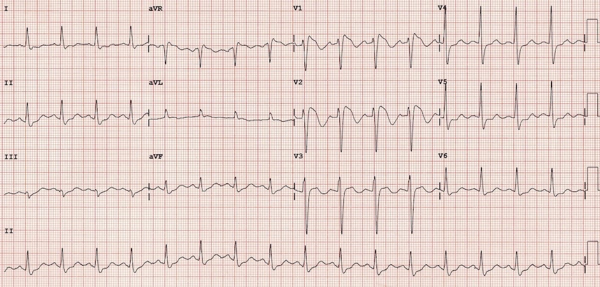
Example 1

RBBB with LAFB.
- Broad QRS > 120ms
- Note the prominent delayed RV conduction, manifested as a tall, broad R wave (R’) best seen in lead V1
- Widened S wave is best appreciated in lead I
- There is appropriate discordance in the right precordial leads with T-wave inversion
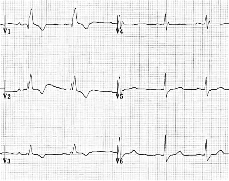
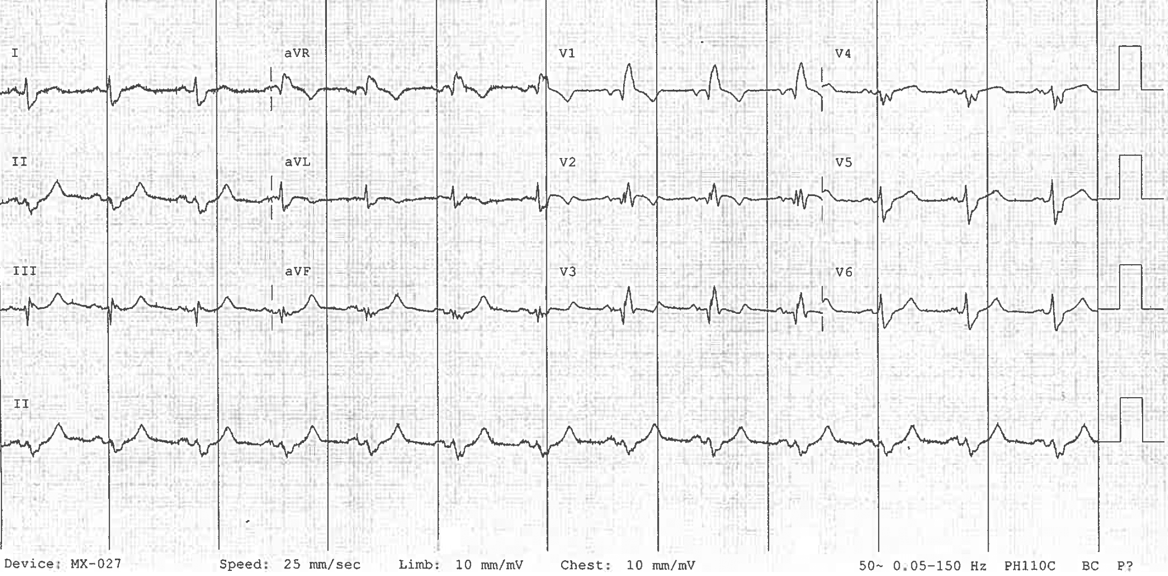
Example 2

Isolated RBBB.
- Typical RSR’ pattern in V1-2
- Widened S waves again demonstrated in lateral leads, especially V4-6
- Appropriate discordance in leads V1-2
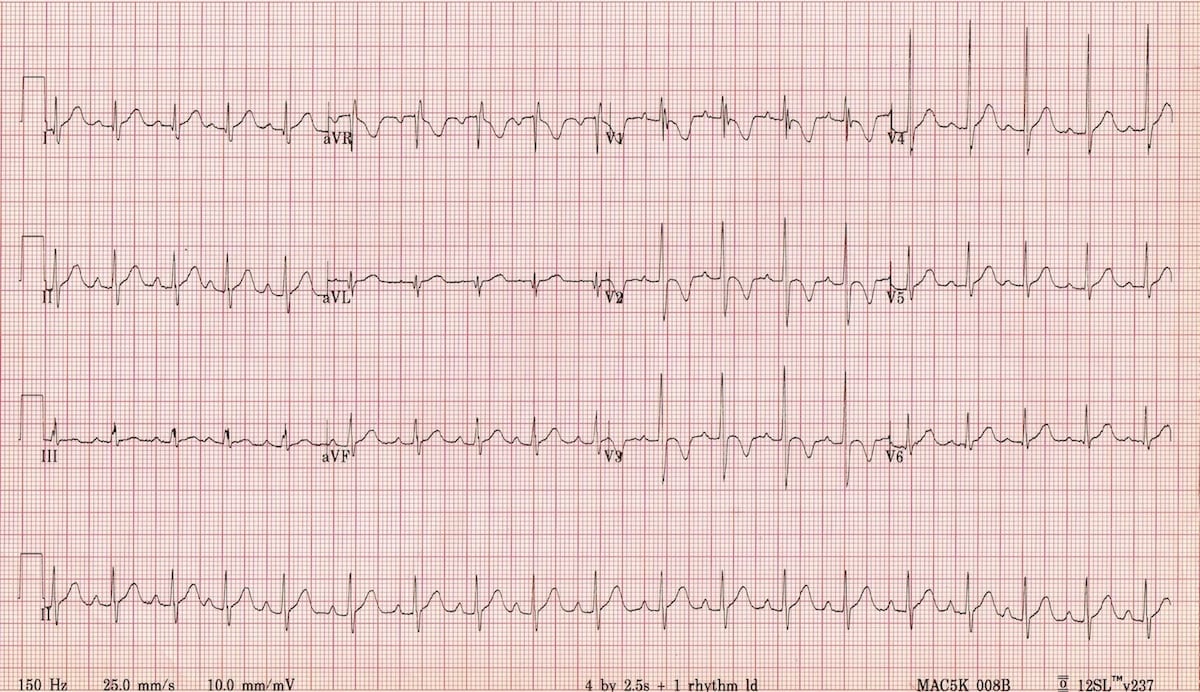
Example 3

Isolated RBBB.
- RSR’ pattern in V1-3
- Lateral S wave changed are not evident here
- Note note normal axis in isolated RBBB
Example 4
RBBB with LAFB in the context of chest pain.
- RBBB is seen with RSR’ pattern in V1-3 and slurred S waves in lateral leads
- There is concordant ST segment changes best seen in V2, and hyper-acute T waves inferiorly. This patient was found to have a 99% proximal LAD occlusion. See OMI: Replacing the STEMI misnomer for further case details
Incomplete RBBB
- Incomplete RBBB is defined as an RSR’ pattern in V1-3 with QRS duration < 120ms.
- It is a normal variant, commonly seen in children (of no clinical significance).
Differential Diagnosis of RBBB
- An RSR’ pattern in V1-3 may also be caused by Brugada syndrome — an ECG pattern associated with malignant ventricular arrhythmias.
Related Topics
- Left bundle branch block LBBB
- Right Bundle Branch Block RBBB
- Left anterior fascicular block LAFB
- Left posterior fascicular block LPFB
- Interventricular Conduction Delay IVCD
- Bifascicular block
- Trifascicular block
- Complete Heart block CHB
- OMI: Replacing the STEMI misnomer
Further reading
- Butterly S, Larsen P. Right Bundle Branch Block. HeartHQ
- Butterly S, Larsen P. Left Bundle Branch Block. HeartHQ
Advanced Reading
Online
- Wiesbauer F, Kühn P. ECG Mastery: Yellow Belt online course. Understand ECG basics. Medmastery
- Wiesbauer F, Kühn P. ECG Mastery: Blue Belt online course: Become an ECG expert. Medmastery
- Kühn P, Houghton A. ECG Mastery: Black Belt Workshop. Advanced ECG interpretation. Medmastery
- Rawshani A. Clinical ECG Interpretation ECG Waves
- Smith SW. Dr Smith’s ECG blog.
- Wiesbauer F. Little Black Book of ECG Secrets. Medmastery PDF
Textbooks
- Zimmerman FH. ECG Core Curriculum. 2023
- Mattu A, Berberian J, Brady WJ. Emergency ECGs: Case-Based Review and Interpretations, 2022
- Straus DG, Schocken DD. Marriott’s Practical Electrocardiography 13e, 2021
- Brady WJ, Lipinski MJ et al. Electrocardiogram in Clinical Medicine. 1e, 2020
- Mattu A, Tabas JA, Brady WJ. Electrocardiography in Emergency, Acute, and Critical Care. 2e, 2019
- Hampton J, Adlam D. The ECG Made Practical 7e, 2019
- Kühn P, Lang C, Wiesbauer F. ECG Mastery: The Simplest Way to Learn the ECG. 2015
- Grauer K. ECG Pocket Brain (Expanded) 6e, 2014
- Surawicz B, Knilans T. Chou’s Electrocardiography in Clinical Practice: Adult and Pediatric 6e, 2008
- Chan TC. ECG in Emergency Medicine and Acute Care 1e, 2004
LITFL Further Reading
- ECG Library Basics – Waves, Intervals, Segments and Clinical Interpretation
- ECG A to Z by diagnosis – ECG interpretation in clinical context
- ECG Exigency and Cardiovascular Curveball – ECG Clinical Cases
- 100 ECG Quiz – Self-assessment tool for examination practice
- ECG Reference SITES and BOOKS – the best of the rest
ECG LIBRARY
Emergency Physician in Prehospital and Retrieval Medicine in Sydney, Australia. He has a passion for ECG interpretation and medical education | ECG Library |

MBBS FACEM DDU (Emergency) CCPU. Emergency Physician in Melbourne, Australia. Co-Ultrasound Lead for Emergency Medicine at The Alfred Hospital. Special interests in diagnostic and procedural ultrasound, medical education, and ECG interpretation. Editor of the LITFL ECG Library.

Quick question for clarification: For a it to be classified as an RSR pattern the S wave needs to be a negative deflection below the isoelectric line right?
So how can the ecg example under “variations” be called a RSR pattern. Isnt it more indicative of a rR pattern?
Keep up the good work – you guys are lifesavers
How about in rsR’ where r wave is greater than the R” wave (Lead V1 and V2). opposite above. is it still called RBBB
Perhaps in a partial RBBB otherwise youre probably seeing an epsilon wave or a normal RSR variant which is not a sign of pathology
[…] Right bundle branch block […]