![]()
Augustus Desiré Waller
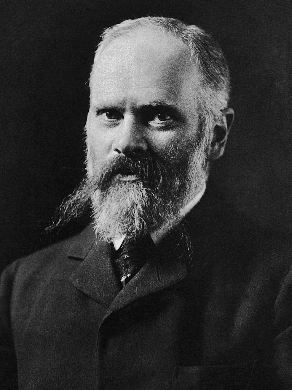
Augustus Desiré Waller (1856-1922) was a Franco-British physiologist.
Waller was best known for recording the first human electrocardiogram (ECG) in 1887. Son of the eminent neurophysiologist Augustus Volney Waller (1816–1870), he trained in medicine at the University of Aberdeen and went on to lecture in physiology at St Mary’s Hospital and the London School of Medicine for Women. His career was characterised by a blend of curiosity-driven experimentation, demonstrative teaching, and a persistent scepticism about the clinical value of his own discoveries.
Waller’s most enduring contribution was the demonstration that the heart’s electrical activity could be detected at the body surface. Using a Lippmann capillary electrometer and custom recording apparatus, he captured rhythmic deflections from human subjects and animals, famously including his dog Jimmy. His public demonstrations were critical in popularising the technique, though Waller himself remained doubtful of its practical medical application. Despite this, his early work directly inspired Willem Einthoven (1860–1927), who refined the method with the string galvanometer and laid the foundations of modern electrocardiography.
Waller proposed the expression for normal systolic duration (1891) which Henry Cuthbert Bazett converted into the Bazett Formula for the measurement of QTc in 1920. His broader physiological research, including his textbook An Introduction to Human Physiology, and his directorship of the University of London’s Physiological Laboratory, reflected his lifelong commitment to integrating experimental science with medical education.
Physiology dominated Waller’s life. He had a home laboratory where his wife, five children and dog Jimmy participated in his experiments. His daughter Mary remembered packing for a trip and her mother asking the children what things they wanted to take, adding ‘but don’t forget, Father has bagged the galvanometer‘.
Biographical Timeline
- Born on July 12, 1856 in Paris, France, son of renowned physiologist Augustus Volney Waller (1816–1870)
- 1870 – Following the death of his father he moved to Aberdeen, Scotland with his mother
- 1878 – Graduated MB, CM from the University of Aberdeen.
- 1881 – Completed MD, University of Aberdeen
- 1882 – Undertook further studies in physiology in Edinburgh, Leipzig (with Carl Ludwig), and Lyon (with Chauveau)
- 1883 – Appointed lecturer in physiology at the London School of Medicine for Women.
- 1884 – Appointed lecturer in physiology at St Mary’s Hospital, Paddington.
- 1885 – Married Alice Palmer, former student and daughter of Sir George Palmer of Huntley & Palmer biscuit fame. Famously greeted at his next St Mary’s lecture by a blackboard inscribed ‘Waller takes the biscuit‘ to which Waller added ‘and the tin as well’.
- 1887 – Recorded the first human electrocardiogram (ECG) using a Lippmann capillary electrometer in May at St Mary’s Hospital.
- 1888 – Publicly demonstrated ECG using saline jars as electrodes; addressed lay audience at St Mary’s Hospital. Elaborated on his studies which also proved that contraction of the heart was not a simultaneous process but showed initiation at the apex and termination at the base.
- 1891 – Published An introduction to Human physiology, a widely used textbook dedicated to his father.
- 1892 – Elected Fellow of the Royal Society (FRS) at age 36.
- 1897 – Appointed Fullerian Professor of Physiology at the Royal Institution.
- 1902 – First Director of the newly founded Physiological Laboratory, University of London, housed at the Imperial Institute.
- 1905 – Consulting physician at the National Hospital for Diseases of the Heart.
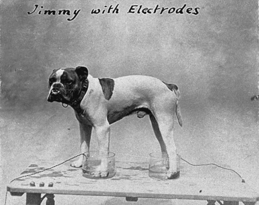
- 1909 – His ECG demonstrations with his bulldog “Jimmy” triggered a parliamentary question under the Cruelty to Animals Act.
- 1913 – Delivered the Oliver-Sharpey Lectures summarising years of research on the heart.
- 1917 – Published a survey of over 2,000 electrocardiograms, and used the term “electro cardiogram” officially.
- Died on March 11, 1922 following a stroke and subsequent haemorrhages
Key Medical Contributions
The electrocardiogram
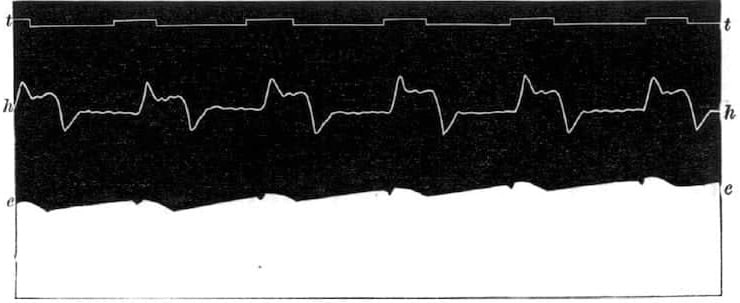
1887 – Recorded the first human electrocardiogram using a capillary electrometer at St Mary’s Hospital, London. Surface electrodes were strapped to the front and back of the chest producing two distorted deflections: ventricular depolarization and repolarization. He attached his equipment to a slowly moving toy train, allowing him to record the heart’s activity in real time.

Waller published initial observations in the Journal of Physiology, illustrating electrical potentials of the human heart and pioneering surface recording of cardiac activity.
If a pair of electrodes (zinc covered by chamois leather and moistened with brine) are strapped to the front and back of the chest, and connected with a Lippmann’s capillary electrometer, the mercury in the latter will be seen to move slightly but sharply at each beat of the heart. If the movements of the column of mercury are photographed on a travelling plate simultaneously with those of an ordinary cardiographic lever a record is obtained
Waller 1887
1888 – Waller delivered a landmark lecture at St Mary’s Hospital Medical School on the electrical activity of the heart in which he described the body-surface recordings of cardiac electrical activity and demonstrated that a human heart acts as an electrical organ. In addition he showed the electrical signal could be transmitted even between individuals holding hands, through the galvanometric circuit.
1889 – Waller published his ‘electro-cardiograph’ results in Philosophical Transactions of the Royal Society including detailed his apparatus and findings, which laid the groundwork for modern electrocardiography.
The contraction of the ventricles is not simultaneous throughout the mass, but traverses it as a wave (at the present stage the direction of the wave of contraction is immaterial). Inequalities of potential, at different parts of the mass, are consequently established at the beginning and at the end of each systole. Or, to reverse the order of statement, the inequalities in question are proof of the passage of a wave of excitation. The distribution of these inequalities of potential is represented diagrammatically:
Waller 1889

1891 – Waller provided a series of values for the duration of mechanical systole with different heart rates. He demonstrated that the period of mechanical contraction (systole) was shortened at faster heart rates. Waller proposed the expression for normal systolic duration, which Henry Cuthbert Bazett converted into the Bazett Formula for the measurement of QTc in 1920.
1909 – Waller often demonstrated the electrocardiograph using his dog “Jimmy” who would patiently stand with paws in glass jars of saline. Jimmy, had the distinction of having had a Parliamentary question asked of him in the House of Commons. Home Secretary Herbert Gladstone defended the dog’s public appearances, explaining that they were painless and Jimmy enjoyed them:
Q. ‘At a conversation of the Royal Society at Burlington House, a bulldog was cruelly treated when a leather strap with sharp nails was wound around his neck and his feet were immersed in glass jars containing salts in solution, and the jars in turn were connected with wires to galvanometers. Such a cruel procedure should surely be dealt with under the “Cruelty to Animals Act” of 1876 ?’
A. ‘The dog in question wore a leather collar ornamented with brass studs, and he was placed to stand in water to which some sodium chloride had been added, or in other words, common salt. If my honourable friend had ever paddled in the sea, he will appreciate fully the sensation obtained thereby from this simple pleasurable experience!’
1912 – Willem Einthoven (1860-1927) credited Waller with coining the term and presenting the first electrocardiogram.
It gives me an especial pleasure to bring to remembrance here that the human EKG was first recorded by a London physiologist, Augustus D. Waller, who also introduced the term “electrocardiogram” into science; and that Dr. Thomas Lewis, whose extensive researches have been crowned with such exceptional success, was the first man in England who applied electrocardiography to clinical investigations. The method of electrocardiography is still a young plant. We may reasonably expect that it will continue to bear good fruit.
Einthoven 1912
1915 – In an informal address at St Mary’s Hospital, Waller reminisced about his pioneer work.
I studied the hearts of all sorts of animals… and one fine day after leading off from the exposed heart of a decapitated cat to study the cardiogram by aid of a Lippman electrometer, it occurred to me that it ought to be possible to use the limbs as electrodes and thus lead off from the heart to the electrometer without exposing the heart, i.e. from the intact and normal organ. Obviously man was the most convenient animal to use so I dipped my right hand and left foot into a couple of basins of salt solution, which were connected with the two poles of the electrometer and at once had the pleasure of seeing the mercury column pulsate with the pulsation of the heart…
This first demonstration was made in St Mary’s laboratory in May 1887 and demonstrated there to many physiologists and among others, to my friend Professor Einthoven of Leiden…During the summer of that year, I made a complete survey of all sorts of leads from the hands and feet and mouth!’
Waller AD. Unpublished address at St Mary’s 1915 (personal communication from Mary Waller to Edwin Besterman, 1951).
1922 – Sir Thomas Lewis (1881-1945) popularized clinical electrocardiography and it is relevant here to quote the words of appreciation of Waller by Lewis:
Waller’s early work on electro-physiology was extensive, thorough, and is well known. He was the first to show that the currents set up by the beating of the human heart can be recorded; he was the first to obtain a human electro-cardiogram; this has been the main though by no means his sole contribution to the science of experimental medicine. The discovery long preceded the introduction of the string galvanometer, and was the more remarkable in that it was accomplished in the eighties.
Thomas Lewis 1922
Controversies
Augustus Desiré Waller used his full name in order to distinguish himself from his eminent father, Augustus Volney Waller (1816–1870), who is remembered for:
- Diapedesis: His thesis involved examination of the frog’s tongue micro-circulation. Waller observed white blood cells (but not red ones) could squeeze between capillary cells.
- Wallerian degeneration of the nerves: Sections of frog’s tongue to show nerves produced ‘necklaces of droplets‘ (degenerating myelin) on one side of the cut – an indication of the direction in which the nerve normally conducted.
Major Publications
- Waller AD. A demonstration on man of electromotive changes accompanying the heart’s beat. Journal of Physiology 1887; 8: 229-234. [First ECG in Man]
- Waller AD. Introductory Address on the Electromotive Properties of the Human Heart. Br Med J. 1888; 2(1449): 751-754
- Waller AD. On the electromotive changes connected with the beat of the mammalian heart, and of the human heart in particular. Philosophical Transactions of the Royal Society B 1889; 180: 169–194.
- Waller AD. An introduction to Human physiology. 1891.
- Waller AD. A lecture on the administration of chloroform to man and to the higher animals: delivered in the Physiological Laboratory of the University of London on October 13th, 1903
- Waller AD. Physiology; the servant of medicine. Chloroform in the laboratory and in the hospital. Univ. of London Press. 1910.
- Waller AD. A contribution to the psychology of logic, considered from a physiological standpoint. University of London Press. 1912.
- Waller AD. The energy value of the ordinary diet of a sedentary worker with and without alcohol. Journal of physiology. 1917; 51: xiii
- Waller AD. A preliminary survey of 2000 Electro Cardiograms. Journal of physiology. 1917; 51: xvii
References
Biography
- Halliburton WD. Augustus Désiré Waller 1856-1922. Royal Society obituary notices 1922
- Augustus Desire Waller (1856-1922) Electrocardiographer. JAMA 1967;202;(2):139-140
- Cope Z. Augustus Desiré Waller (1856-1922). Med Hist. 1973 Oct;17(4):380-5.
- Sykes AH. A. D. Waller: a biographical note. St Marys Hosp Gaz. 1985;91(3):19-20.
- Besterman E, Creese R. Waller–pioneer of electrocardiography. Br Heart J. 1979 Jul;42(1):61-4.
- Acierno LJ. Augustus Desiré Waller. Clin Cardiol. 2000 Apr;23(4):307-9.
- Sykes AH. Servants of Medicine: Augustus Waller—father and son physiologists. J R Soc Med. 2005; 98(4): 185–186
- Bibliography. Waller, Augustus Desiré 1856-1922. WorldCat Identities
Eponymous terms
- Einthoven W. The different forms of the human electrocardiogram and their signification. Lancet 1912; 179(4622): 853-861
- Waller MD. Discovery of the Electrocardiogram. Br Med J. 1950; 1(4660): 1008–1009.
- Besterman E. Waller before Einthoven in the clinical development of noninvasive ECG. Am Heart J. 1982 Apr;103(4 Pt 1):572
- Sykes AH. A D Waller and the electrocardiogram, 1887. Br Med J (Clin Res Ed). 1987; 294(6584): 1396-8
- Lüderitz B. Augustus Desiré Waller (1856-1922)–the first to record the electrical activity of the human heart. J Interv Card Electrophysiol. 2003 Aug;9(1):59-60.
- Cadogan M. History of the Electrocardiogram. LITFL 2020
- Morris T. The first electrocardiogram (ECG). 2021
Eponym
the person behind the name

MBBS (Hons) FCEM. Emergency Consultant at St Mary's Hospital, Imperial College Healthcare NHS Trust
BA MA (Oxon) MBChB (Edin) FACEM FFSEM. Emergency physician, Sir Charles Gairdner Hospital. Passion for rugby; medical history; medical education; and asynchronous learning #FOAMed evangelist. Co-founder and CTO of Life in the Fast lane | On Call: Principles and Protocol 4e| Eponyms | Books |