
![]()
Bashed, Blind and Bulging
aka Ophthalmology Befuddler 033
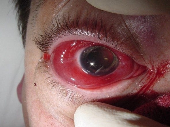
A 35 year-old martial artist presents with loss of vision in his right eye after being on the wrong end of a spinning back fist.
Examination of the right eye reveals:
- He is unable to detect light when the eyelids are passively opened.
- There is a relative afferent pupillary defect affecting the right eye.
- Extraocular movements are markedly reduced.
- Tonometry reveals an intraocular pressure of 45 mmHg.
Questions
Q1. What is shown and what is the likely diagnosis?
Answer and interpretation
The right eye appears proptosed with a dilated pupil, extensive subconjunctival hemorrhage and bloody chemosis, as well evidence of periorbital swelling and hematoma.
The likely underlying diagnosis is:
Retrobulbar hemorrhage due to trauma, resulting in acute orbital compartment syndrome.
Hemorrhage into the potential space within the rigid orbit and around the eye transmits pressure onto the optic nerve. Acute orbital compartment syndrome occurs when this results in a compressive optic neuropathy.
The main differentials are:
- ruptured globe: proptosis and raised intraocular pressure are not consistent with this
- orbital blowout fracture: raised intraocular pressure, RAPD and decreased visual acuity are not seen in this condition unless there is coexistent retrobulbar hemorrhage.
Q2. What are the clinical features of this condition on history?
Answer and interpretation
History:
- Symptoms: pain, decreased vision, inability to open the eyelids due to severe swelling.
- Cause: history of trauma or surgery to the eye or orbit, or retrobulbar injection.
- Risk factors for spontaneous hemorrhage — bleeding disorder, anticoagulants and anti-platelet drugs, pregnancy.
Q3. What are the clinical features of this condition on examination?
Answer and interpretation
Examination:
- Visual acuity and visual fields: decreased with dyschromatopsia (signs of optic neuropathy)
- External exam: Proptosis with resistance to retropulsion, diffuse subconjunctival hemorrhage, tight eyelids (rock hard) with echymosis and chemosis.
- Extraocular eye movements: limited extraocular motility
- Pupils: RAPD.
- Tonometry: increased intraocular pressure (IOP)
- Funduscopy: papilloedema from compressive optic neuropathy may be present, retinal artery or vein occlusion.
Q4. What investigation should be performed and what are the typical findings?
Answer and interpretation
Retrobulbar haemorrhage with acute orbital compartment syndrome is primarily a clinical diagnosis.
CT orbit (axial and coronal views) can confirm the diagnosis — but if vision is threatened treat first!
Usual findings on CT:
- diffuse, increased reticular pattern of the intraconal orbital fat rather than a discrete hematoma.
- teardrop or tenting sign is ominous — it occurs when the optic nerve is at maximum stretch and distorts the back of the globe into a teardrop shape.
Q5. What is the treatment of this condition?
Answer and interpretation
Admit to hospital and treat with aggressive decompression.
Therapy depends on whether there is compressive optic neuropathy or severely raised IOP:
- Evidence of optic neuropathy or severely raised IOP (>40 mmHg): lateral canthotomy and cantholysis should be performed immediately (ideally by an ophthalmologist); use procedural sedation in the ED if it does not cause a delay.
- No evidence of optic neuropathy but IOP is raised (e.g. >30 mmHg): treat with agents used to lower IOP (e.g. topical timolol, acetazolamide, mannitol; see acute glaucoma).
McInnes and Howes suggest the DIP-A CONE-G mnemonic for remembering the indications and contra-indications for this procedure:
Primary indications:
- Decreased visual acuity
- Intraocular pressure > 40 mm Hg
- Proptosis
Secondary indications:
- Afferent pupillary defect
- Cherry red macula
- Ophthalmoplegia
- Nerve head pallor
- Eye pain
Contraindications:
- Globe rupture
Q6. How is a lateral canthotomy/ cantholysis performed?
Answer and interpretation
The main steps in emergency canthotomy/ cantholysis are:
- use local anesthetic but warn the patient that they may feel pain
- Perform the canthotomy:
- place the scissors across the lateral canthus and incise the canthus full thickness
- Perform cantholysis:
- Grasp the lateral lower eyelid with toothed forcepsPull the lower eyelid anteriorlyPoint the scissors toward the patient’s nose, place the blades either side of the lateral canthal tendon, and cut.
Cantholysis is done more by feel than by visual identification of landmarks — the lower eyelid will come completely away from the globe once the tendon has been completely severed.
Good overview from Ballard et al: Emergency lateral canthotomy and cantholysis: a simple procedure to preserve vision from sight threatening orbital hemorrhage.
Emergency lateral canthotomy and cantholysis: a simple procedure to preserve vision from sight threatening orbital hemorrhage.
References
- Johnson D, Schweitzer K, Sharma S. Ophthaproblem: Can you identify this condition? Retrobulbar hemorrhage. Can Fam Physician. 2009 Jun;55(6):605, 607. PMC2694083.
- Ballard SR, Enzenauer RW, O’Donnell T, Fleming JC, Risk G, Waite AN. Emergency lateral canthotomy and cantholysis: a simple procedure to preserve vision from sight threatening orbital hemorrhage. J Spec Oper Med. 2009 Summer;9(3):26-32. [PMID 19739474]
- McInnes G, Howes DW. Lateral canthotomy and cantholysis: a simple, vision-saving procedure. CJEM. 2002 Jan;4(1):49-52. PMID: 17637149.
- Perry M. Acute proptosis in trauma: retrobulbar hemorrhage or orbital compartment syndrome — does it really matter? J Oral Maxillofac Surg. 2008 Sep;66(9):1913-20. PMID: 18718400.
- Ehlers JP, Shah CP, Fenton GL, Hoskins EN. The Wills Eye Manual: Office and Emergency Room Diagnosis and Treatment of Eye Disease Lippincott Williams & Wilkins
- NSW Statewide Opthalmology Service. Eye Emergency Manual — An illustrated Guide. [Free PDF]
OPHTHALMOLOGY BEFUDDLER
Chris is an Intensivist and ECMO specialist at The Alfred ICU, where he is Deputy Director (Education). He is a Clinical Adjunct Associate Professor at Monash University, the Lead for the Clinician Educator Incubator programme, and a CICM First Part Examiner.
He is an internationally recognised Clinician Educator with a passion for helping clinicians learn and for improving the clinical performance of individuals and collectives. He was one of the founders of the FOAM movement (Free Open-Access Medical education) has been recognised for his contributions to education with awards from ANZICS, ANZAHPE, and ACEM.
His one great achievement is being the father of three amazing children.
On Bluesky, he is @precordialthump.bsky.social and on the site that Elon has screwed up, he is @precordialthump.
| INTENSIVE | RAGE | Resuscitology | SMACC