![]()
Henri Huchard
Henri Huchard (1844–1910), French cardiologist at Necker, defined “cardio-arterial” disease, described Huchard’s sign, and helped shape early hypertension care.

![]()
Henri Huchard (1844–1910), French cardiologist at Necker, defined “cardio-arterial” disease, described Huchard’s sign, and helped shape early hypertension care.

Rigler notch sign: Indentation in the border of a solid lung mass (thought to represent a feeding vessel) suggestive of a bronchial carcinoma
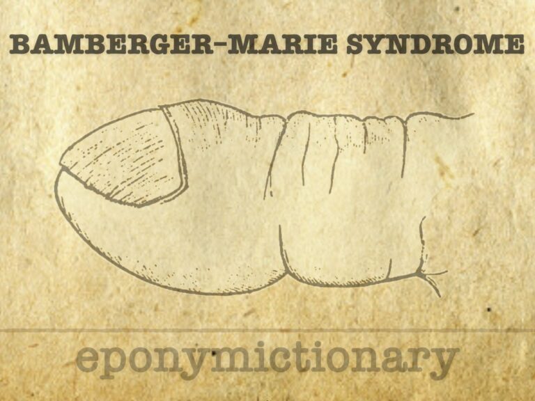
Bamberger–Marie syndrome (hypertrophic pulmonary osteoarthropathy): clubbing, bone periostosis, and joint effusions—historically recognised as a paraneoplastic syndrome linked to lung disease.
Eugen von Bamberger (1858–1921), Austrian internist; co-described Marie–Bamberger syndrome (hypertrophic pulmonary osteoarthropathy); pioneer of clinical diagnostics.
Louis Virgil Hamman (1877–1946), Johns Hopkins physician and diagnostician, described Hamman’s sign, Hamman syndrome, and Hamman-Rich syndrome

EGPA (Churg–Strauss syndrome): rare ANCA-associated vasculitis with asthma, eosinophilia, and systemic granulomatous inflammation of small vessels
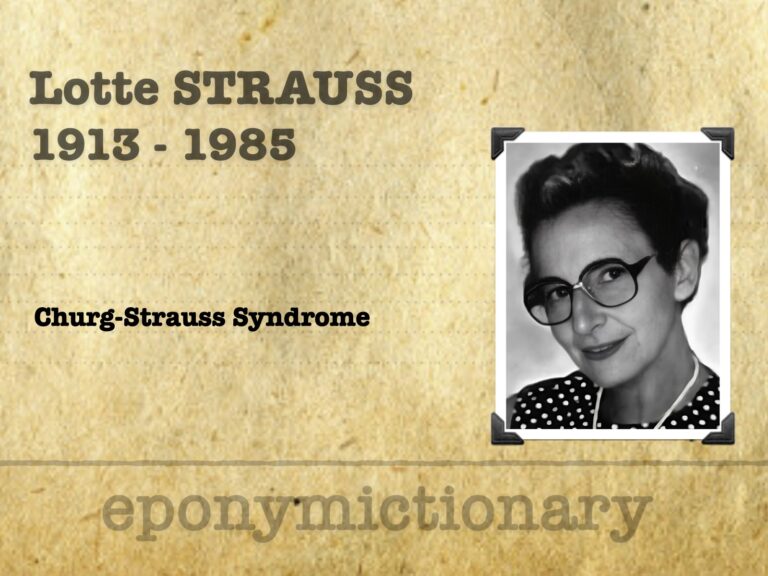
Lotte Strauss (1913–1985), pioneer in paediatric and perinatal pathology, co-described Churg–Strauss syndrome and helped found the Society for Pediatric Pathology
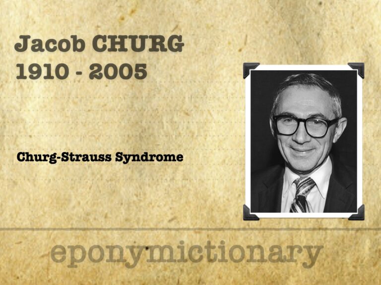
Jacob Churg (1910–2005), pioneering pathologist, co-described Churg–Strauss syndrome and transformed renal pathology through biopsy-based diagnostics
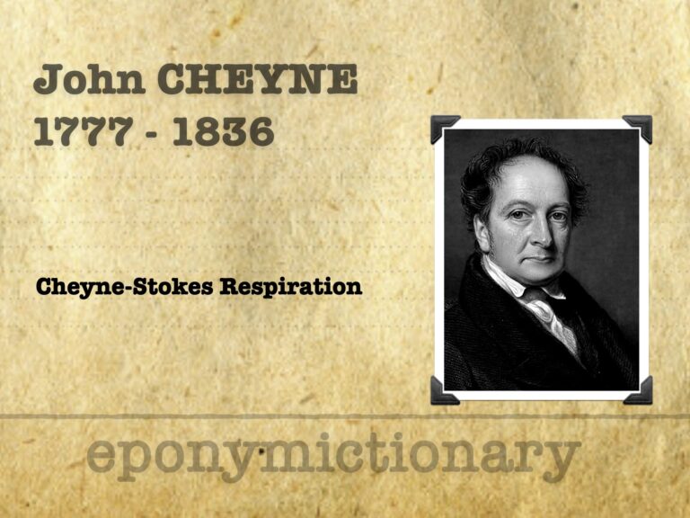
John Cheyne (1777–1836), Irish physician, co-described Cheyne-Stokes respiration, advanced clinical neurology, and linked pupils to brain injury
Cheyne-Stokes respiration is a cyclical breathing pattern of apnoea and hyperpnoea, seen in heart failure, brain injury, and end-of-life settings.

Henry Khunrath Pancoast (1875 – 1939) was an American radiologist. The Pancoast tumour and Pancoast syndrome is named after him

Pancoast Tumour is a primary bronchogenic carcinoma which arises in the apex of the lung at the superior pulmonary sulcus.