![]()
CXR Case 064
A 56 year old man presents with increasing right sided shoulder pain, unresponsive to low dose opiates.
Describe and interpret this CXR
CHEST X-RAY INTERPRETATION
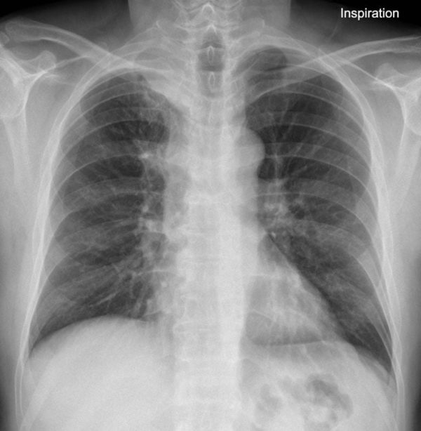
There is increased soft tissue shadowing in the right apex and airspace shadowing in the right upper lobe.
The right hilum is raised. Otherwise lung fields are clear.
No obvious rib destruction around the lesion.
CLINICAL CORRELATION
This is a Pancoast tumour.
Pancoast tumour – a primary lung cancer in the apex of the lung that classically invades the surrounding soft tissues causing often very significant pain with musculoskeletal and sometimes brachial plexus invasion.
CLINICAL PEARLS
Henry Pancoast – the first Professor of Radiology in the United States – first described this appearance in 1924.
Remember that a Pancoast Tumor is associated with Pancoast Syndrome in patients with shoulder pain, C8-T2 radiculopathy and ipsilateral Horner syndrome. (Johan Friedrich Horner was a Swiss opthalmologist!)
TOP 150 CXR SERIES
![]()
![]()
![]()
Prof Fraser Brims Curtin Medical School, acute and respiratory medicine specialist, immediate care in sport doc, ex-Royal Navy, academic| Top 100 CXR | Google Scholar | ICIS Course ANZ