
![]()
ECG Case 058
Elderly patient presenting with chest pain. Interpret the ECG.
Describe and interpret this ECG
ECG ANSWER and INTERPRETATION
Main Abnormalities
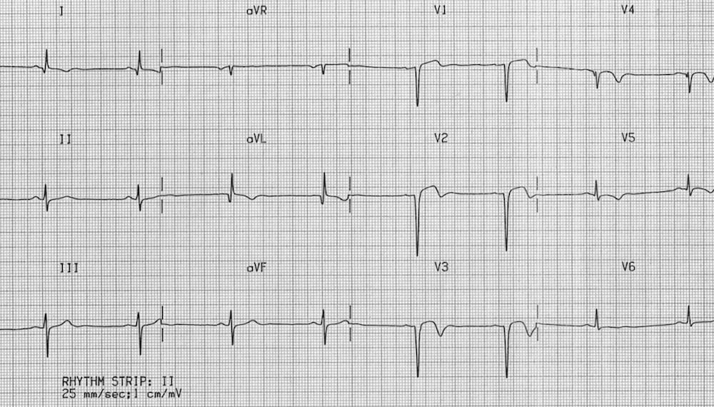
This ECG shows changes consistent with an old anterolateral infarction — the so-called left ventricular aneurysm pattern.
- ST elevation in V1-3 associated with deep Q waves (= LV aneurysm morphology).
- Pathological Q waves also seen in I, aVL and V4.
- Poor R wave progression (= R wave height < 3 mm in V3).
- Biphasic / inverted T waves in V1-5, I and aVL.
CLINICAL PEARLS
The LV aneurysm pattern refers to the combination of residual ST elevation, deep Q waves and inverted or biphasic T waves seen in patients following an acute myocardial infarction. This ECG pattern is associated with transmural scarring and paradoxical movement of the LV on wall on echocardiography.
Around 60% of patients with anterior STEMI develop some degree of chronic ST elevation on their ECG, which can cause diagnostic confusion.
If these patients present with chest pain, the safest approach is to take serial ECGs looking for signs of evolving STEMI such as evolving ST elevation or pseudo-normalisation of T waves.
References
Further Reading
- Wiesbauer F, Kühn P. ECG Mastery: Yellow Belt online course. Understand ECG basics. Medmastery
- Wiesbauer F, Kühn P. ECG Mastery: Blue Belt online course: Become an ECG expert. Medmastery
- Kühn P, Houghton A. ECG Mastery: Black Belt Workshop. Advanced ECG interpretation. Medmastery
- Rawshani A. Clinical ECG Interpretation ECG Waves
- Smith SW. Dr Smith’s ECG blog.
- Wiesbauer F. Little Black Book of ECG Secrets. Medmastery PDF
TOP 100 ECG Series
Emergency Physician in Prehospital and Retrieval Medicine in Sydney, Australia. He has a passion for ECG interpretation and medical education | ECG Library |

If the patient’s pain had resolved; could the vignette and ECG be interpreted as Wellens?
Wellens is defined as without precordial Q waves (https://litfl.com/wellens-syndrome-ecg-library/). The QS waves here are pretty characteristic of LV aneurysm pattern/old infarct. On serial ECGs, you would also expect evolution of the precordial biphasic T waves in Wellens given biphasic waves are an early finding.