![]()
ECG Case 059
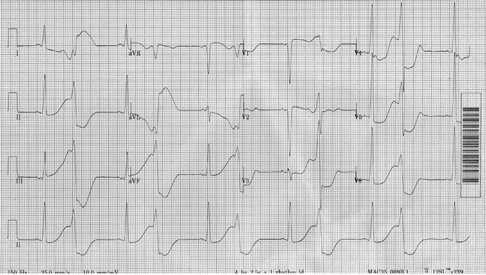
57-year old man with ROSC following VF arrest. Interpret the ECG.
Describe and interpret this ECG
ECG ANSWER and INTERPRETATION
Main Abnormalities
- Sinus rhythm with frequent ventricular ectopics in a pattern of ventricular bigeminy.
- Grossly prolonged QT interval (> 600 ms).
- “R on T” phenomenon is present, with each VEB falling on the end of the T wave — this ECG pattern is very high risk for deterioration to torsades de pointes and ventricular fibrillation.
- Relatively short PR interval and possible delta waves (leads I, II, V6) are suggestive — but not diagnostic — of WPW syndrome.
- Voltage criteria for left ventricular hypertrophy are present in multiple leads.
This patient had suffered a cardiac arrest in the context of severe hypertrophic cardiomyopathy and long QT syndrome.
(NB. ~1/3 of patients with HOCM will have some evidence of WPW on their ECG).
Can you guess what happened next?
The patient had a further TdP cardiac arrest!
This was treated with IV magnesium and potassium, with restoration of sinus rhythm.
This interesting case is discussed in Cardiovascular Curveball 003
References
Further Reading
- Wiesbauer F, Kühn P. ECG Mastery: Yellow Belt online course. Understand ECG basics. Medmastery
- Wiesbauer F, Kühn P. ECG Mastery: Blue Belt online course: Become an ECG expert. Medmastery
- Kühn P, Houghton A. ECG Mastery: Black Belt Workshop. Advanced ECG interpretation. Medmastery
- Rawshani A. Clinical ECG Interpretation ECG Waves
- Smith SW. Dr Smith’s ECG blog.
- Wiesbauer F. Little Black Book of ECG Secrets. Medmastery PDF
TOP 100 ECG Series
Emergency Physician in Prehospital and Retrieval Medicine in Sydney, Australia. He has a passion for ECG interpretation and medical education | ECG Library |