
![]()
Q Wave
The Q Wave
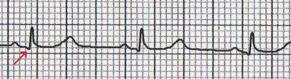
A Q wave is any negative deflection that precedes an R wave
- The Q wave represents the normal left-to-right depolarisation of the interventricular septum
- Small ‘septal’ Q waves are typically seen in the left-sided leads (I, aVL, V5 and V6)
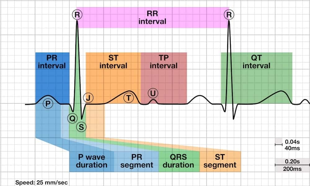
Q waves in context
Q waves in different leads
- Small Q waves are normal in most leads
- Deeper Q waves (>2 mm) may be seen in leads III and aVR as a normal variant
- Under normal circumstances, Q waves are not seen in the right-sided leads (V1-3)
Pathological Q Waves
Q waves are considered pathological if:
- > 40 ms (1 mm) wide
- > 2 mm deep
- > 25% of depth of QRS complex
- Seen in leads V1-3
Pathological Q waves usually indicate current or prior myocardial infarction.
Differential Diagnosis
- Myocardial infarction
- Cardiomyopathies — Hypertrophic (HCM), infiltrative myocardial disease
- Rotation of the heart — Extreme clockwise or counter-clockwise rotation
- Lead placement errors — e.g. upper limb leads placed on lower limbs
Loss of normal Q waves
- The absence of small septal Q waves in leads V5-6 should be considered abnormal.
- Absent Q waves in V5-6 is most commonly due to LBBB.
ECG Examples
Example 1
- Inferior Q waves (II, III, aVF) with ST elevation due to acute MI
Example 2
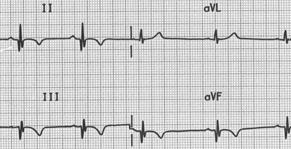
- Inferior Q waves (II, III, aVF) with T-wave inversion due to previous MI
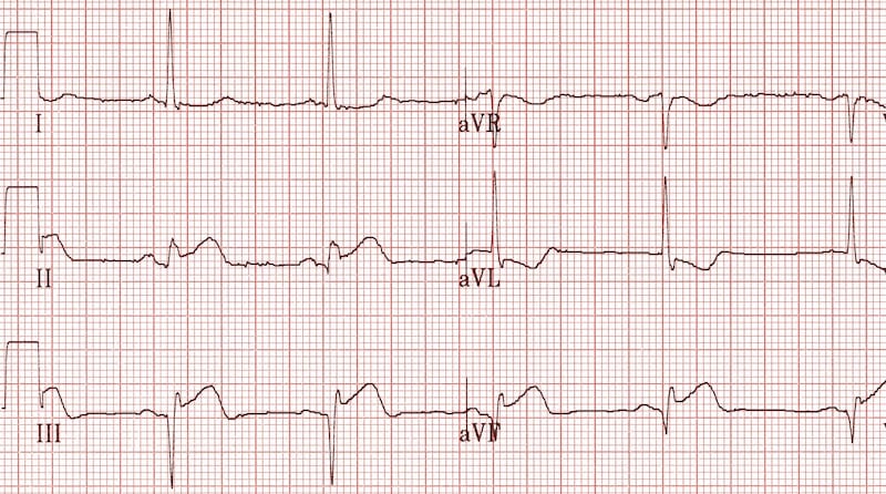
Example 3
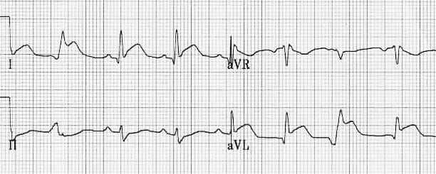
- Lateral Q waves (I, aVL) with ST elevation due to acute MI
Example 4
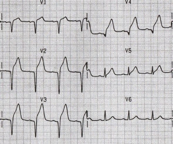
- Anterior Q waves (V1-4) with ST elevation due to acute MI
Example 5
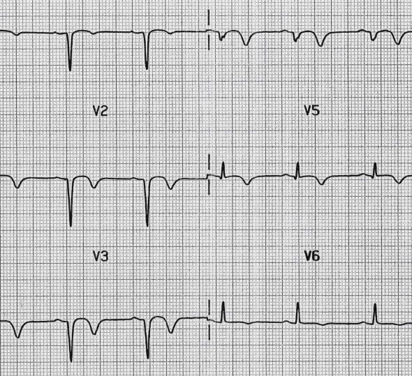
- Anterior Q waves (V1-4) with T-wave inversion due to recent MI
ECG Library Basics
Advanced Reading
Online
- Wiesbauer F, Kühn P. ECG Mastery: Yellow Belt online course. Understand ECG basics. Medmastery
- Wiesbauer F, Kühn P. ECG Mastery: Blue Belt online course: Become an ECG expert. Medmastery
- Kühn P, Houghton A. ECG Mastery: Black Belt Workshop. Advanced ECG interpretation. Medmastery
- Rawshani A. Clinical ECG Interpretation ECG Waves
- Smith SW. Dr Smith’s ECG blog.
- Wiesbauer F. Little Black Book of ECG Secrets. Medmastery PDF
Textbooks
- Zimmerman FH. ECG Core Curriculum. 2023
- Mattu A, Berberian J, Brady WJ. Emergency ECGs: Case-Based Review and Interpretations, 2022
- Straus DG, Schocken DD. Marriott’s Practical Electrocardiography 13e, 2021
- Brady WJ, Lipinski MJ et al. Electrocardiogram in Clinical Medicine. 1e, 2020
- Mattu A, Tabas JA, Brady WJ. Electrocardiography in Emergency, Acute, and Critical Care. 2e, 2019
- Hampton J, Adlam D. The ECG Made Practical 7e, 2019
- Kühn P, Lang C, Wiesbauer F. ECG Mastery: The Simplest Way to Learn the ECG. 2015
- Grauer K. ECG Pocket Brain (Expanded) 6e, 2014
- Surawicz B, Knilans T. Chou’s Electrocardiography in Clinical Practice: Adult and Pediatric 6e, 2008
- Chan TC. ECG in Emergency Medicine and Acute Care 1e, 2004
LITFL Further Reading
- ECG Library Basics – Waves, Intervals, Segments and Clinical Interpretation
- ECG A to Z by diagnosis – ECG interpretation in clinical context
- ECG Exigency and Cardiovascular Curveball – ECG Clinical Cases
- 100 ECG Quiz – Self-assessment tool for examination practice
- ECG Reference SITES and BOOKS – the best of the rest
ECG LIBRARY
Emergency Physician in Prehospital and Retrieval Medicine in Sydney, Australia. He has a passion for ECG interpretation and medical education | ECG Library |

MBBS DDU (Emergency) CCPU. Adult/Paediatric Emergency Medicine Advanced Trainee in Melbourne, Australia. Special interests in diagnostic and procedural ultrasound, medical education, and ECG interpretation. Co-creator of the LITFL ECG Library. Twitter: @rob_buttner

Thankyou for this Dr Burns,
I wonder what your thoughts are on finding inferior Q waves in a patient with an indeterminate axis? Should we still expect the small inferior R waves seen in a superior axis?
Thank you for this concise but informative post.
Just a question: would that be safe to say that in example 4 and 5, there are only Q waves in Leads V1 to V4, and no R wave, no S wave, then we are onto ST elevation?
Yes correct. Otherwise known as poor R wave progression, we see complete loss of R waves which is common in acute infarction.
[…] https://litfl.com/q-wave-ecg-library/ […]
Why we considered Q wave and T waves inversion as recent MI in Example 5 but in Example 2 considered as previous?