

Pediatric CXR Cases 007
Pediatric Emergency Medicine Chest X-ray interpretation with Jennifer Potter and Nicholena Richardson


Pediatric Emergency Medicine Chest X-ray interpretation with Jennifer Potter and Nicholena Richardson
November Adult Emergency Medicine Chest X-ray interpretation with Alyssa Thomas, MD and Claire Milam, MD
October Pediatric Emergency Medicine Chest X-ray interpretation with Jennifer Potter and Nicholena Richardson from EMGuideWire
June Pediatric Emergency Medicine Chest X-ray interpretation with Jennifer Potter and Nicholena Richardson from EMGuideWire
Pediatric Emergency Medicine Radiology Topics, monthly educational, self-guided slides were first published on EMGuideWire.com and peer reviewed by Professor Gibbs and Sean Fox, MD
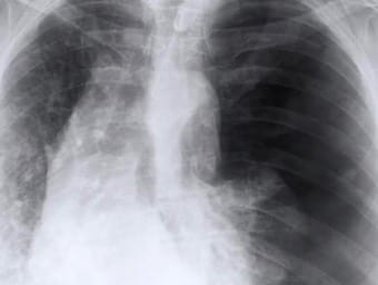
ATMIST handover - 28 y/o male, injured 25 mins ago, penetrating chest trauma, Asherman seal anterior chest, RR 35, deteriorating, high flow O2 administered.

Peter James Kerley first described horizontal lines that he postulated to be peri-vascular lymphatics in patients with mitral stenosis and left ventricular failure
Hilar enlargement reflects one of 4 types of processes: Lymphadenopathy and tumors; Pulmonary venous hypertension; Pulmonary arterial hypertension; or Increased pulmonary blood flow
Additional reading from Normal CXRs; Eric Strong Interpretation series; the DRABCDE approach; CXR for the OSCE and of course the Top 150 CXR to try your luck!
Labelled normal anatomy chest X-ray to assist in interpretation review
Chest X-Rays (CXR) are routine investigation in clinical practice and consequently it is important for medical students and clinician’s alike to know how to interpret them. There are many approaches to CXR interpretation, each trying to ensure that key abnormalities are identified and no area is overlooked.
A 69 yo lady presents with a fall, productive cough, fever and dyspnoea. LITFL Top 150 CXR