
![]()
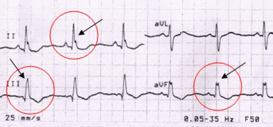
Crochetage sign
Notch near the apex of the R wave of inferior leads (II, III, aVF) seen in a large proportion of patients with an ostium secundum atrial septal defect (ASD), the most common form of ASD
1958 – First described by Toscano et al, the electrophysiological mechanism behind this ECG change remains unknown
1996 – Heller et al described the crochetage pattern of the R wave in inferior limb leads as frequent in patients with atrial septal defect, correlating with shunt severity and being independent of the right bundle branch block pattern.
The retrospective case study observed this pattern in at least one inferior lead of 73% of patients with a secundum ASD; and 36% of patients with ventricular septal defect (VSD), compared with 7% of the general population. Specificity for the diagnosis of ASD increased to 92-100% when associated with an incomplete RBBB pattern, or when present in all three inferior leads
In ASD, the incidence of Crochetage sign increases with larger anatomic defect or greater left-to-right shunt
1998 – Ay et al concluded that the finding of an ECG crochetage pattern may help to identify stroke patients with PFO, may help to streamline their diagnostic workup, and may warrant future studies to determine its value in stratifying stroke risk in patients with PFO.
The sensitivity (36%) and specificity (91%) of the crochetage pattern for diagnosis of PFO in cryptogenic stroke with had a positive predictive value was 77%.
2018 – Lei Shen found 28% of patients with ASD had the characteristic R wave notching in all three inferior leads compared to just 2% of age matched controls
ECG features of secundum ASD
- Characteristic R wave notching in inferior leads (Crochetage sign)
- Slight right axis deviation (RAD)
- Voltage evidence of right ventricular hypertrophy (RVH), often in the form of “incomplete” right bundle branch block (RBBB)
ECG Examples of Crochetage sign
Example 1
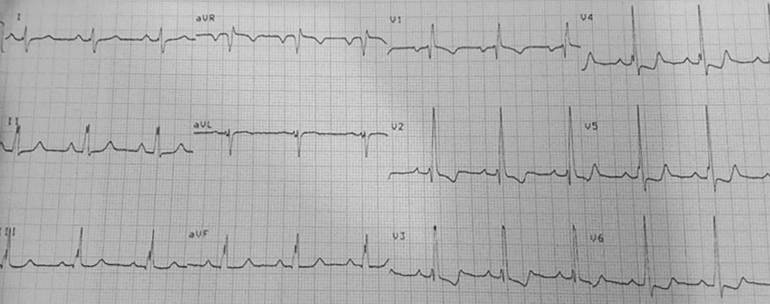
- Right axis deviation
- Incomplete RBBB pattern
- Crochetage sign in leads II, III and aVF
This patient was found to have a 20mm ostium secundum ASD. There was resolution of crochetage sign following operative repair
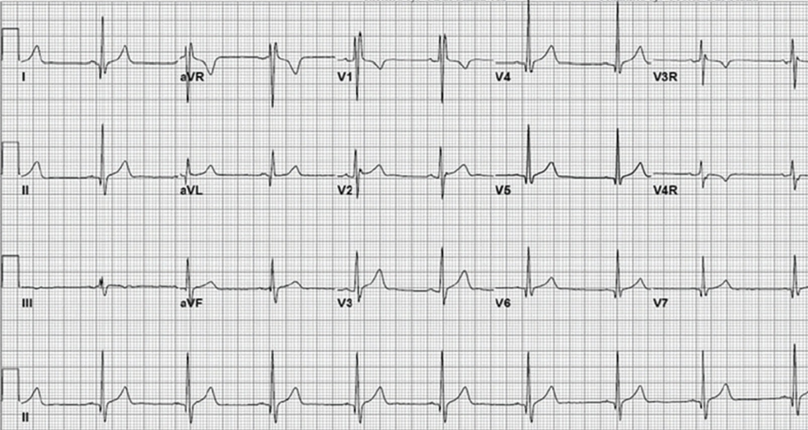
Example 2

Example 3
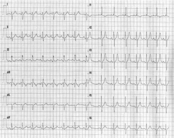
- Right axis deviation
- Crochetage sign in leads II, III and aVF
References
- Toscano Barboza E, Brandenburg RO, Swan HJ. Atrial septal defect: the electrocardiogram and its hemodynamic correlation in 100 proved cases. Am J Cardiol. 1958;2:698–713.
- Davia JE, Cheitlin MD, Bedynek JL. Sinus venosus atrial septal defect: analysis of fifty cases. Am Heart J. 1973; 85: 177–185.
- Fournier A, Young ML, Garcia OL, Tamer DF, Wolff GS. Electrophysiologic cardiac function before and after surgery in children with atrioventricular canal. Am J Cardiol. 1986; 57: 1137–1141.
- Heller J et al. “Crochetage” (Notch) on R wave in inferior limb leads: A new independent electrocardiographic sign of atrial septal defect. J Am Coll Cardiol. 1996 Mar, 27 (4) 877–882
- Ay H, Buonanno FS, Abraham SA, Kistler JP, Koroshetz WJ. An electrocardiographic criterion for diagnosis of patent foramen ovale associated with ischemic stroke. Stroke. 1998 Jul;29(7):1393-7
- Jost CH, Connolly HM, Danielson GK, Bailey KR, Schaff HV, Shen WK, Warnes CA, Seward JB, Puga FJ, Tajik AJ. Sinus venosus atrial septal defect: long-term postoperative outcome for 115 patients. Circulation. 2005; 112: 1953–1958.
- Webb G, Gatzoulis MA. Atrial septal defects in the adult: recent progress and overview. Circulation. 2006;114:1645–1653
Advanced Reading
Online
- Wiesbauer F, Kühn P. ECG Mastery: Yellow Belt online course. Understand ECG basics. Medmastery
- Wiesbauer F, Kühn P. ECG Mastery: Blue Belt online course: Become an ECG expert. Medmastery
- Kühn P, Houghton A. ECG Mastery: Black Belt Workshop. Advanced ECG interpretation. Medmastery
- Rawshani A. Clinical ECG Interpretation ECG Waves
- Smith SW. Dr Smith’s ECG blog.
- Wiesbauer F. Little Black Book of ECG Secrets. Medmastery PDF
Textbooks
- Zimmerman FH. ECG Core Curriculum. 2023
- Mattu A, Berberian J, Brady WJ. Emergency ECGs: Case-Based Review and Interpretations, 2022
- Straus DG, Schocken DD. Marriott’s Practical Electrocardiography 13e, 2021
- Brady WJ, Lipinski MJ et al. Electrocardiogram in Clinical Medicine. 1e, 2020
- Mattu A, Tabas JA, Brady WJ. Electrocardiography in Emergency, Acute, and Critical Care. 2e, 2019
- Hampton J, Adlam D. The ECG Made Practical 7e, 2019
- Kühn P, Lang C, Wiesbauer F. ECG Mastery: The Simplest Way to Learn the ECG. 2015
- Grauer K. ECG Pocket Brain (Expanded) 6e, 2014
- Surawicz B, Knilans T. Chou’s Electrocardiography in Clinical Practice: Adult and Pediatric 6e, 2008
- Chan TC. ECG in Emergency Medicine and Acute Care 1e, 2004
LITFL Further Reading
- ECG Library Basics – Waves, Intervals, Segments and Clinical Interpretation
- ECG A to Z by diagnosis – ECG interpretation in clinical context
- ECG Exigency and Cardiovascular Curveball – ECG Clinical Cases
- 100 ECG Quiz – Self-assessment tool for examination practice
- ECG Reference SITES and BOOKS – the best of the rest
ECG LIBRARY

MBBS DDU (Emergency) CCPU. Adult/Paediatric Emergency Medicine Advanced Trainee in Melbourne, Australia. Special interests in diagnostic and procedural ultrasound, medical education, and ECG interpretation. Co-creator of the LITFL ECG Library. Twitter: @rob_buttner