![]()
Abdominal CT: Common Terms
Describing pathology like a radiologist
Next, let’s cover some essential terminology that is used to describe pathology seen on CT images. Here we review common terms such as fat stranding, fluid and thickened as related to the CT scan.
What is fat stranding?
Many structures in the abdomen are surrounded by fat. Normally, the fat appears dark with a similar density throughout the abdominal cavity. Fat stranding is a term that is almost synonymous with inflammation on a CT exam and can be seen with a large variety of pathologies.
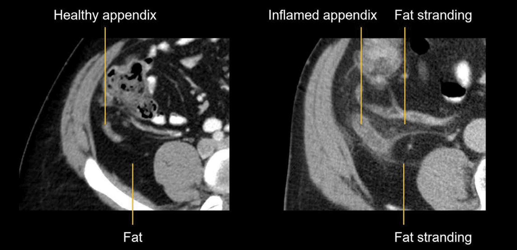
Example 1: Appendicits
Below is a CT image (left) demonstrating a normal appendix with surrounding fat.
When the appendix becomes inflamed (CT image on the right), the fat surrounding it no longer appears dark. Instead, it becomes brighter and may have wavy lines through it. This is called fat stranding and indicates inflammation.
Example 2: Sigmoid inflammation
Below is a CT image (left) demonstrating a normal sigmoid colon with dark fat surrounding it, similar in appearance to the fat of the body wall. However in the CT image on the right is a sigmoid colon with inflammation. Again you can see that the fat surrounding it is not dark, but instead has stranding and a general hazy appearance.
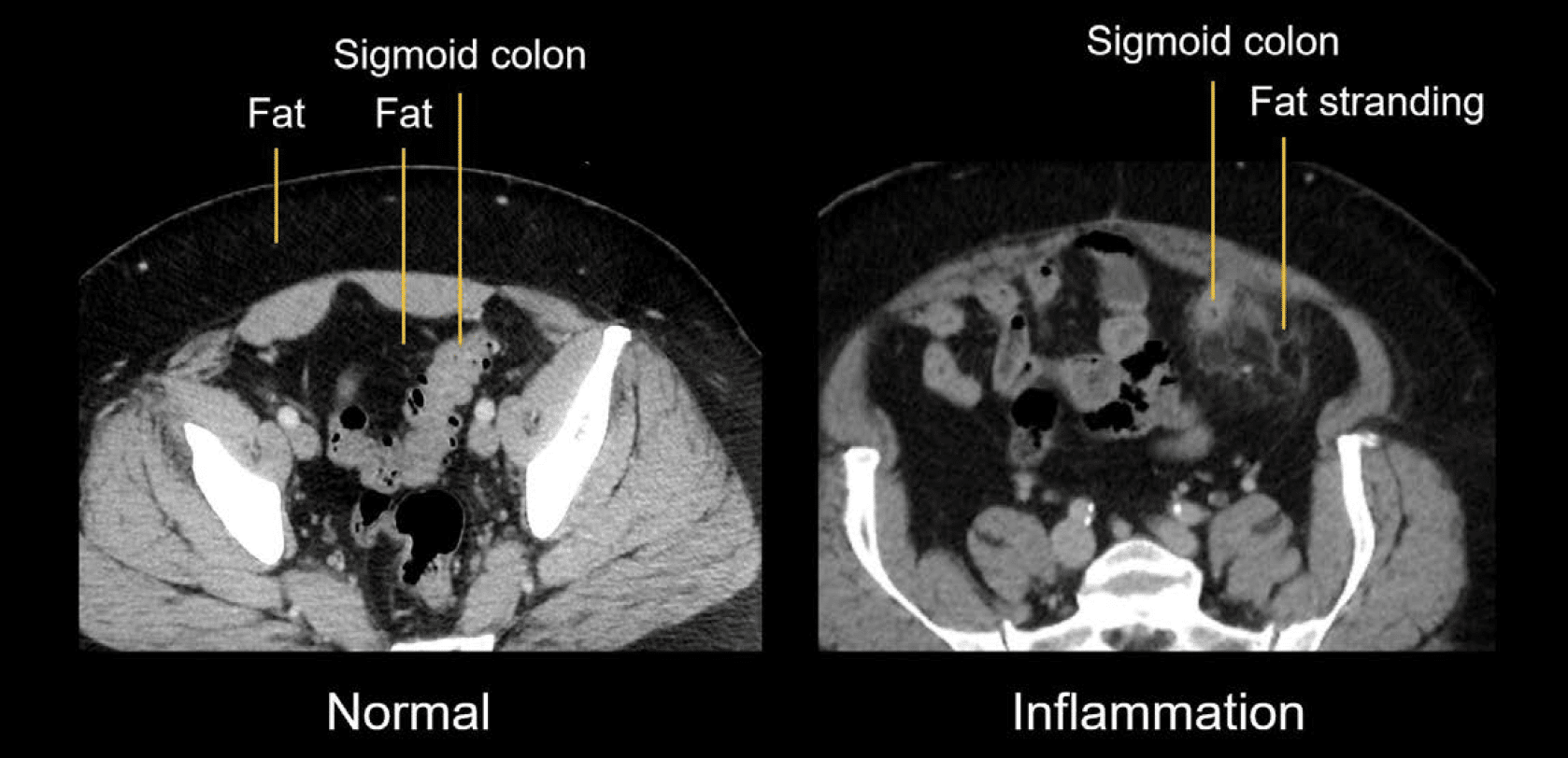
Over time, your eyes will become trained to evaluate fat in a CT image, as it serves as a messenger letting you know that the structure next to it is having trouble.
What does fluid look like on CT?
Getting used to recognising fluid in the abdominal cavity takes time, but it is an important finding to make. Fluid is brighter on a CT image than fat, but less bright than the organs or bones.
Normally, the abdominal cavity does not have free fluid. However, patients with liver disease, for example, can develop fluid in the abdominal cavity, which replaces the fat and makes it look more grey. We can see fluid in four places in the image below.
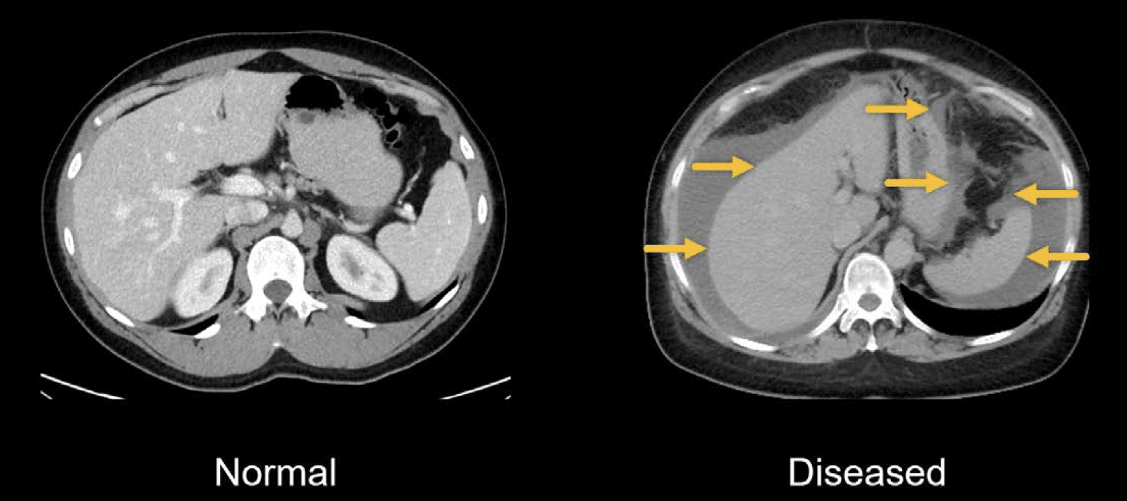
Note: Fluid can be denser and appear brighter if it has blood or cancer cells within it.
What does thickened mean?
When you describe something as thickened on CT images, you are usually comparing:
- What you observe on the images in relation to your concept of normal.
- The structure you observe in relation to neighbouring structures.
Of course, making these comparisons gets easier with experience.
Below is an example of a thickened stomach wall, which is in the gastric antrum to be exact. When viewing an image, you can compare what you are seeing to normal stomach examples or surrounding structures. For example, the wall of the gastric body of this patient is normal, which helps to emphasize the thickening in the antrum.

This is an edited excerpt from the Medmastery course Abdominal CT Essentials by Michael P. Hartung, MD. Acknowledgement and attribution to Medmastery for providing course transcripts.
- Hartung MP. Abdominal CT: Common Pathologies. Medmastery
- Hartung MP. Abdominal CT: Essentials. Medmastery
- Hartung MP. Abdomen CT: Trauma. Medmastery
References
- Top 100 CT scan quiz. LITFL
Radiology Library: Abdominal CT Basics
- Hartung MP. What is the role of Abdominal CT? LITFL
- Hartung MP. Abdominal CT: Basics. LITFL
- Hartung MP. Abdominal CT: Common Terms. LITFL
- Hartung MP. Abdominal CT: Planes. LITFL
- Hartung MP. Abdominal CT: Measuring attenuation. LITFL
- Hartung MP. Abdominal CT: Windows settings (basics). LITFL
- Hartung MP. Abdominal CT: Windows settings (advanced). LITFL
Abdominal CT interpretation

Assistant Professor of Abdominal Imaging and Intervention at the University of Wisconsin Madison School of Medicine and Public Health. Interests include resident and medical student education, incorporating the latest technology for teaching radiology. I am also active as a volunteer teleradiologist for hospitals in Peru and Kenya. | Medmastery | Radiopaedia | Website | Twitter | LinkedIn | Scopus

MBChB (hons), BMedSci - University of Edinburgh. Living the good life in emergency medicine down under. Interested in medical imaging and physiology. Love hiking, cycling and the great outdoors.