![]()
Abdominal CT: Windows advanced
How do I adjust the window settings to evaluate ANY structure?
In the PACS windows basics post, we reviewed the generic window settings which can be applied to shift the grey scale range, enabling you to see structures of different densities better. Here, we are going to dive deeper into those window settings and how they relate to attenuation values.
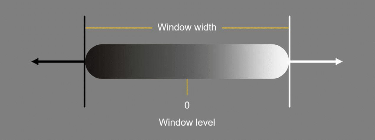
Window settings refers to two key parameters:
- Window width: which determines the range of grey scale values that the image will display
- Window level: which defines the centre point of that range
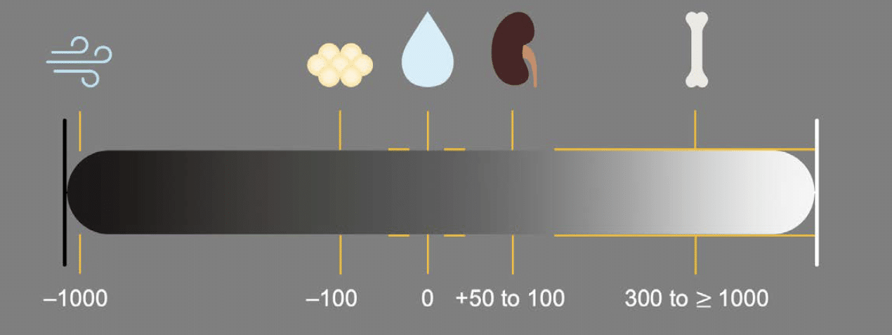
Both settings are measured in Hounsfield units. These values correspond to how a tissue blocks x-rays, called attenuation. By convention water is 0 HU, air is –1000 HU, fat is around –100 HU, soft tissue is about 50–100 HU, and bone can range from 300 HU to more than 1000 HU depending on how dense it is.
Example 1: Soft tissue window
Let’s apply this to the soft tissue window, where the window width is 400 and the window level, or center point, is 50. This means that the gray scale is spread out over 400 HU total, ranging from –150 to 250 HU. Everything with an HU value below –150 will appear black, and everything with an HU value greater than 250 will appear white.

These window settings concentrate the greyscale differences over soft tissue structures. Here, the centre point is 50, right in the soft tissue range.
The specific settings in the soft tissue window help us to see relatively subtle but important differences in enhancement or densities of structures like the liver, spleen, pancreas, and kidneys, which helps us diagnose different diseases.
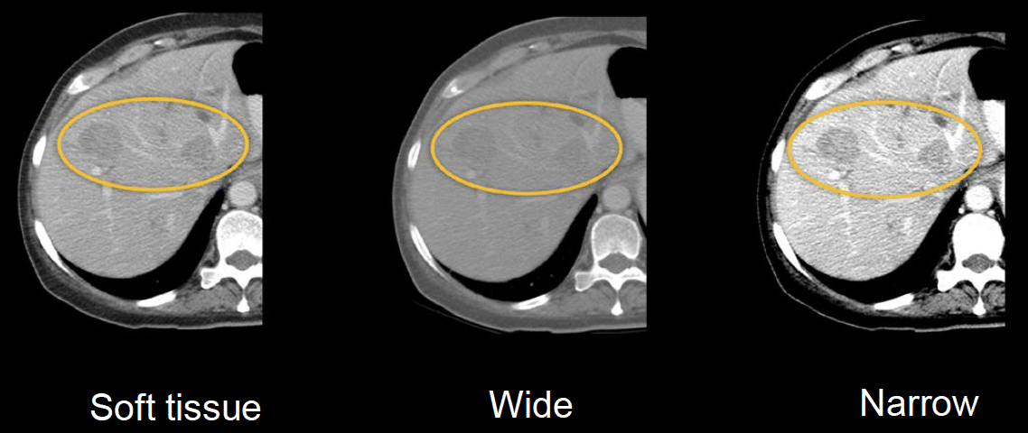
Example 2: Liver metastases and the liver window
There are instances when it can be helpful to further adjust the window width to draw out these subtle differences. The CT images below are from a patient with liver masses that are relatively hard to see on the soft tissue window.
- If we widen the window width, the transition from dark to light structures happens more gradually. This obscures differences in soft tissue and makes them blend together with less definition.
- If we narrow the window, the changes in the grey scale values occur more abruptly, which emphasises subtle differences and makes it easier to find subtle abnormalities in the soft tissues
In fact, a narrower window is so commonly used for viewing the liver that it is referred to as the liver window.
Adjusting window level
Changing the window level, or centre point, essentially makes the whole image brighter or darker. The biggest difference can be seen between the lung and bone windows.
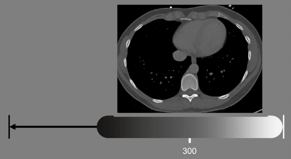
Example 3: Bone
When viewing bones, the midpoint is higher because you want the centre of your grey scale values to optimize your ability to evaluate denser bone. In the case shown next, the level is about 300 HU.
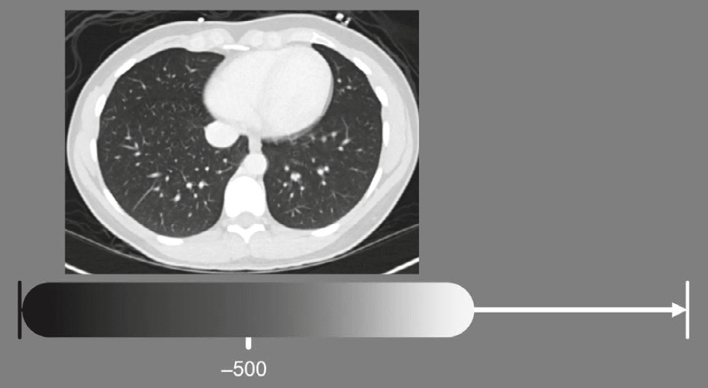
Example 4: Lung
When viewing the lungs, the centre point must be quite low, around –500, because you want to centre your grey scale values to optimize your ability to see air-filled structures. Remember, pure air is –1000 HU by convention.
Example 5: bowel perforation
Bone, soft tissue, and lung windows of the abdomen in a patient with a bowel perforation with air is leaking into the abdominal cavity. We can see the free air most confidently using the lung window because the level is centred on air, making the difference between air (black) and soft tissue (white) the most dramatic.
After identifying free air, evaluating the bowel in the soft tissue window is helpful when trying to find the exact location of the perforation.

Summary
There are three most common and useful window settings for your image review:
- Soft tissue
- Bone
- Lung
Each setting has a range of grey scale values it displays called the window width, and a centre point called the window level. You should practice using various window settings to help you get the most from your CT images.
This is an edited excerpt from the Medmastery course Abdominal CT Essentials by Michael P. Hartung, MD. Acknowledgement and attribution to Medmastery for providing course transcripts.
- Hartung MP. Abdominal CT: Common Pathologies. Medmastery
- Hartung MP. Abdominal CT: Essentials. Medmastery
- Hartung MP. Abdomen CT: Trauma. Medmastery
References
- Top 100 CT scan quiz. LITFL
Radiology Library: Abdominal CT Basics
- Hartung MP. What is the role of Abdominal CT? LITFL
- Hartung MP. Abdominal CT: Basics. LITFL
- Hartung MP. Abdominal CT: Common Terms. LITFL
- Hartung MP. Abdominal CT: Planes. LITFL
- Hartung MP. Abdominal CT: Measuring attenuation. LITFL
- Hartung MP. Abdominal CT: Windows settings (basics). LITFL
- Hartung MP. Abdominal CT: Windows settings (advanced). LITFL
Abdominal CT interpretation

Assistant Professor of Abdominal Imaging and Intervention at the University of Wisconsin Madison School of Medicine and Public Health. Interests include resident and medical student education, incorporating the latest technology for teaching radiology. I am also active as a volunteer teleradiologist for hospitals in Peru and Kenya. | Medmastery | Radiopaedia | Website | Twitter | LinkedIn | Scopus

MBChB (hons), BMedSci - University of Edinburgh. Living the good life in emergency medicine down under. Interested in medical imaging and physiology. Love hiking, cycling and the great outdoors.