
![]()
Abdominal CT: Planes
Distinguishing imaging planes
Firstly, we review how computed tomography (CT) is different from X-ray, and then cover computed tomography planes.
How is CT different from X-ray?
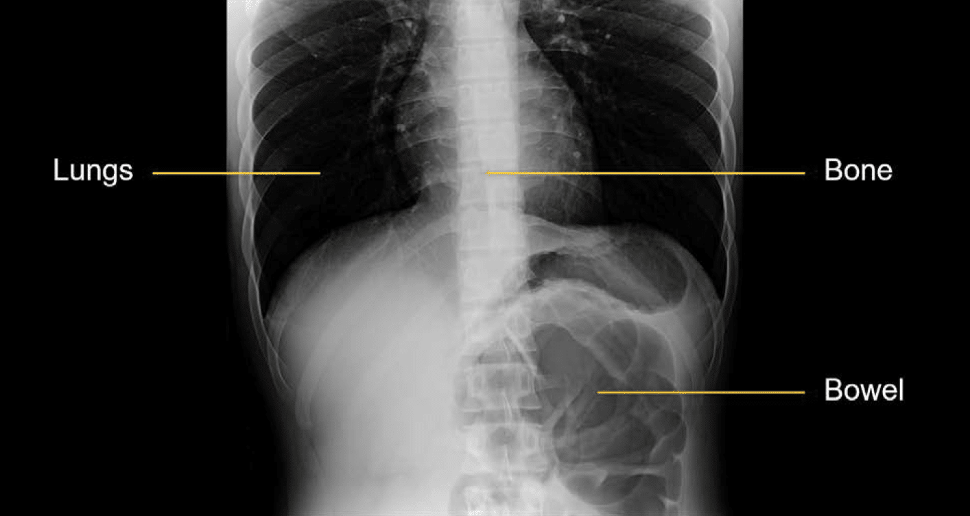
X-rays are obtained with the patient in between the x-ray generator and the x-ray detector. Unlike a CT scanner which can be manipulated to view images in multiple planes, an x-ray can only capture an image in a single plane. The process is similar to taking a picture with a camera, with the obvious difference that the x-ray camera allows you to see what is under the skin.

The x-ray detector records how the x-rays are blocked by structures in the body and this is used to generate an image. Dense structures like bone block x-rays and look bright or white on the images. Air-filled structures like lung or bowel look darker because they do not block many x-rays.
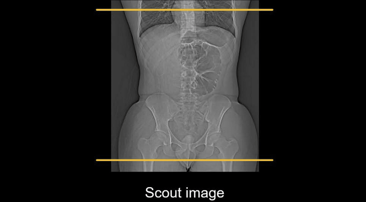
A CT scan starts by taking an image similar to an x-ray called the scout or localiser image. This image is used to plan the CT exam. For example, in an abdominopelvic CT, the scout image is used to plan the scan from just above the bottom of the lung to just below the pubic symphysis.
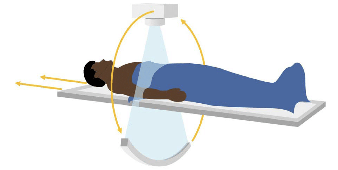
During a CT scan, the tube and detectors spin very quickly around the patient while continuously taking pictures as the patient passes through the scanner. This provides information about how the body blocks x-rays from every angle. This extra information is used to create 3-dimensional images which allow you to scroll through the body slice by slice and see very detailed anatomy.
What are the CT planes?
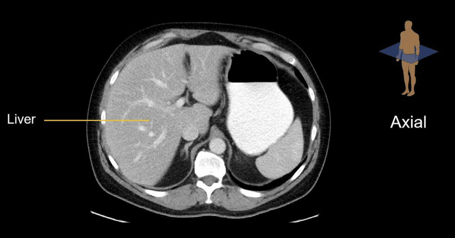
Once the patient images are obtained, they are reconstructed first in the axial plane, which reflects how the images were taken. The axial plane can be thought of as slices through the patient
Axial plane
The axial plane is the one that radiologists use the most, but it is not the most intuitive. Let’s face it, we don’t think about people in slices, and the anatomic right and left are reversed on the image. For example, in this image, the liver is found on the left side of the image, but anatomically it is located on the right.
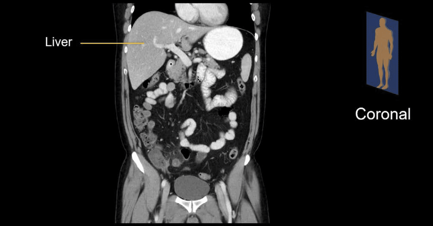
Coronal plane
The most intuitive view for beginners is the coronal plane. This plane reflects how we look at the patient as they stand in front of us or lie on the examination table. Even though the liver is still on the left, we can imagine ourselves looking directly at the patient, and thus we expect to see a mirror-image of their anatomy. This view also provides the most direct comparison to abdominal x-ray.
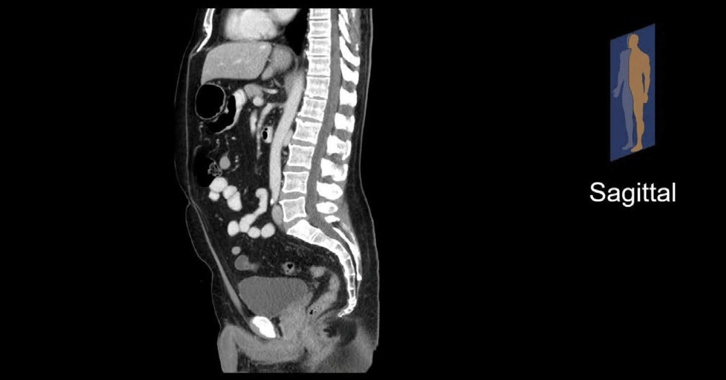
Sagittal plane
The sagittal plane is the final plane that is generated. For the abdominal organs and bowel, the sagittal view is largely reserved for problem solving any abnormalities that may have been identified on the other views. However, it is very useful for reviewing the spine, as this view allows you to see how the vertebral bodies stack up with each other.
Over time you will become accustomed to how each organ and structure appears in each view and the pros and cons of each imaging plane.
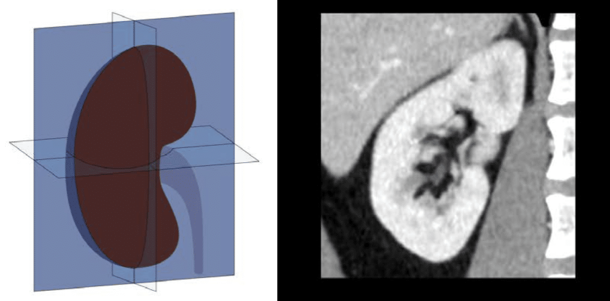
Worked example: the kidney
As an example, let’s look at the kidney in all three planes.
The axial plane is your starting point for evaluating the kidneys. But, as you scroll through slice by slice, it can be hard to get a sense of the overall shape and enhancement of the kidney in this view.

The coronal plane is the most intuitive view of the kidney because it shows you the entire kidney in its long axis allowing you to take in the size, shape, and overall enhancement. It also allows you to compare the left and right sides in a single image.
The sagittal plane also displays the long axis of the kidney, but from a different angle. This view can help you evaluate complex pathology that may be difficult to see on axial or coronal images.

This is an edited excerpt from the Medmastery course Abdominal CT Essentials by Michael P. Hartung, MD. Acknowledgement and attribution to Medmastery for providing course transcripts.
- Hartung MP. Abdominal CT: Common Pathologies. Medmastery
- Hartung MP. Abdominal CT: Essentials. Medmastery
- Hartung MP. Abdomen CT: Trauma. Medmastery
References
- Top 100 CT scan quiz. LITFL
Radiology Library: Abdominal CT Basics
- Hartung MP. What is the role of Abdominal CT? LITFL
- Hartung MP. Abdominal CT: Basics. LITFL
- Hartung MP. Abdominal CT: Common Terms. LITFL
- Hartung MP. Abdominal CT: Planes. LITFL
- Hartung MP. Abdominal CT: Measuring attenuation. LITFL
- Hartung MP. Abdominal CT: Windows settings (basics). LITFL
- Hartung MP. Abdominal CT: Windows settings (advanced). LITFL
Abdominal CT interpretation

Assistant Professor of Abdominal Imaging and Intervention at the University of Wisconsin Madison School of Medicine and Public Health. Interests include resident and medical student education, incorporating the latest technology for teaching radiology. I am also active as a volunteer teleradiologist for hospitals in Peru and Kenya. | Medmastery | Radiopaedia | Website | Twitter | LinkedIn | Scopus

MBChB (hons), BMedSci - University of Edinburgh. Living the good life in emergency medicine down under. Interested in medical imaging and physiology. Love hiking, cycling and the great outdoors.
