![]()
Abdominal CT: Basics
How to speak like a radiologist
Learning abdominal computed tomography (CT) can feel a little overwhelming, almost like speaking a different language. As you review and interpret more and more CT images, you will become increasingly comfortable with the terminology and more familiar with what normal and abnormal findings look like.
Here we review the basics of attenuation, enhancement and orientation.
What is Attenuation?
Attenuation determines what appears bright and dark on a CT image. CT images are shown in a range of grey scale values from black and shades of grey to white. These values reflect the density of the structure being viewed.
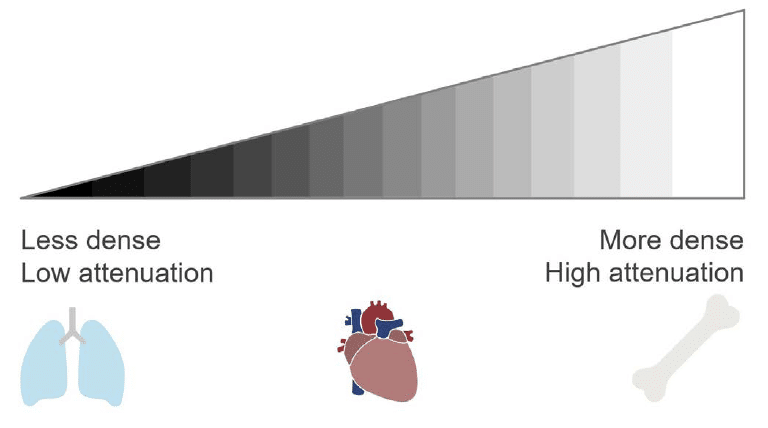
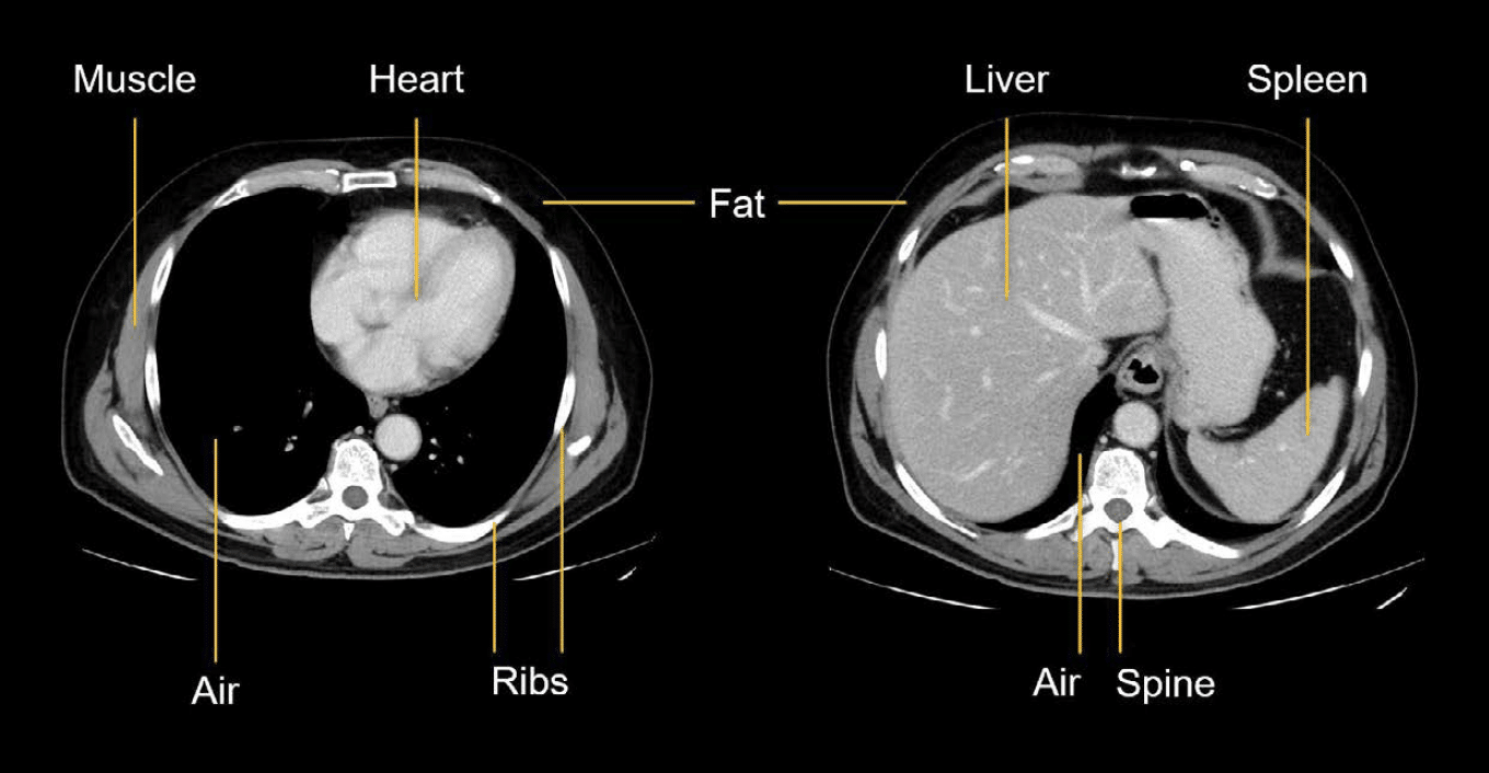
When looking at structures in the abdomen, darker areas mean fewer x-rays are blocked, so the tissue is less dense. For example, air and fat will appear black. Conversely, white areas mean that many of the x-rays are blocked because the tissue is denser—like bone. The range of gray values covers the different densities of fluid and soft tissue we associate with muscle and organs.
However, there is more information in a single image than can be displayed in gray scale. For instance, take a look at the CT images below. We can see the lower chest in the image on the left, but we can’t really see the lungs because they are almost completely black except for a few dots from the pulmonary vessels.
Similar to editing a photo by changing the exposure or contrast, we can change the way the CT image is displayed so that we can see the lung tissue. This is called changing the window. By adjusting the contrast, the soft tissues appear white, as shown in the image on the right.

What is enhancement?
Now that we have talked about what makes an image bright or dark, let’s discuss enhancement, and how intravenous (IV) contrast can be used to improve the details in CT images.
IV contrast mixes with and is carried through the blood. When it enters a structure, the structure will appear brighter on CT; it enhances. IV contrast will make the structure look brighter than it would without contrast.
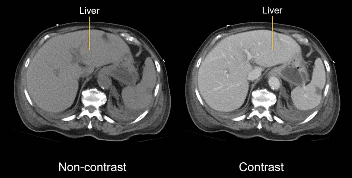
You can compare IV contrast to turning on a light in a dark room. When the light is off, you can see the basic shape of things, but without detail. When you turn the light on with contrast, things brighten and you can see everything more clearly, including the inside of organs.

Assessing enhancement can help you identify abnormalities. For example, inflammation often increases enhancement making the structure brighter than normal. When blood flow is blocked, as in ischemia, the structure will enhance less, appearing less bright than normal.
Below is an example of kidneys, without and with contrast. Notice how the enhanced kidneys appear brighter. In the image with contrast, you may have noticed that the upper part of the left kidney appears darker. This difference in enhancement is caused by blocked blood flow, which could only be detected with the use of contrast.

What is orientation?
Finally, we have terms to orient ourselves when describing findings.

Proximal and distal
Proximal and distal are used to establish a point of reference along the length of a structure. Proximal refers to the beginning of the reference point and distal refers to the end of it.
Proximal and distal are particularly relevant for tubular structures like the bowel and blood vessels. For example, if there is a mass blocking the end of the large intestine, you can say that the distal bowel is obstructed and, as a result of the obstruction, the proximal bowel is dilated.
Medial / lateral, anterior / posterior, and superior / inferior
These terms are helpful when describing the relationship of different structures or the location of areas of concern.
- Medial describes ‘close to the midline or middle’, whilst lateral defines an area toward the side/outside, away from the midline.
- Anterior is toward the front, and posterior is toward the back.
- Superior (cranial) is toward the head, and inferior (caudal) is toward the feet, or away from the head.
This is an edited excerpt from the Medmastery course Abdominal CT Essentials by Michael P. Hartung, MD. Acknowledgement and attribution to Medmastery for providing course transcripts.
- Hartung MP. Abdominal CT: Common Pathologies. Medmastery
- Hartung MP. Abdominal CT: Essentials. Medmastery
- Hartung MP. Abdomen CT: Trauma. Medmastery
References
- Top 100 CT scan quiz. LITFL
Radiology Library: Abdominal CT Basics
- Hartung MP. What is the role of Abdominal CT? LITFL
- Hartung MP. Abdominal CT: Basics. LITFL
- Hartung MP. Abdominal CT: Common Terms. LITFL
- Hartung MP. Abdominal CT: Planes. LITFL
- Hartung MP. Abdominal CT: Measuring attenuation. LITFL
- Hartung MP. Abdominal CT: Windows settings (basics). LITFL
- Hartung MP. Abdominal CT: Windows settings (advanced). LITFL
Abdominal CT interpretation

Assistant Professor of Abdominal Imaging and Intervention at the University of Wisconsin Madison School of Medicine and Public Health. Interests include resident and medical student education, incorporating the latest technology for teaching radiology. I am also active as a volunteer teleradiologist for hospitals in Peru and Kenya. | Medmastery | Radiopaedia | Website | Twitter | LinkedIn | Scopus

MBChB (hons), BMedSci - University of Edinburgh. Living the good life in emergency medicine down under. Interested in medical imaging and physiology. Love hiking, cycling and the great outdoors.