
![]()
ECG Case 041
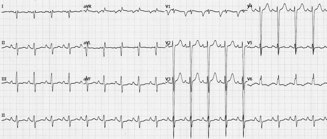
70-year old patient presenting with acute pulmonary oedema. Describe the ECG
Describe and interpret this ECG
ECG ANSWER and INTERPRETATION
This is an ECG example of dilated cardiomyopathy demonstrating signs of enlargement of all four cardiac chambers:
- There is marked LVH with very deep S waves in V2-4.
- Right axis deviation suggests associated RV enlargement (= biventricular enlargement).
- Evidence of left atrial enlargement (deep, wide terminal portion of the P wave in V1).
- Peaked P waves in lead II suggestive of right atrial enlargement (~ 2.5mm in height).
This patient had four-chamber dilatation on echocardiography with severe congestive cardiac failure (awaiting cardiac transplantation).
References
Further Reading
- Wiesbauer F, Kühn P. ECG Mastery: Yellow Belt online course. Understand ECG basics. Medmastery
- Wiesbauer F, Kühn P. ECG Mastery: Blue Belt online course: Become an ECG expert. Medmastery
- Kühn P, Houghton A. ECG Mastery: Black Belt Workshop. Advanced ECG interpretation. Medmastery
- Rawshani A. Clinical ECG Interpretation ECG Waves
- Smith SW. Dr Smith’s ECG blog.
- Wiesbauer F. Little Black Book of ECG Secrets. Medmastery PDF
TOP 100 ECG Series
Emergency Physician in Prehospital and Retrieval Medicine in Sydney, Australia. He has a passion for ECG interpretation and medical education | ECG Library |

It’s possible see still two signs of RAE: Sodi-Pallares sign – qR (in this case qRs) in V1 (normal is rS); and Peñaloza-Tranchesi sign – fast change QRS shape V1 to V2, little in the first and normal in the last one.