
![]()
ECG Case 069
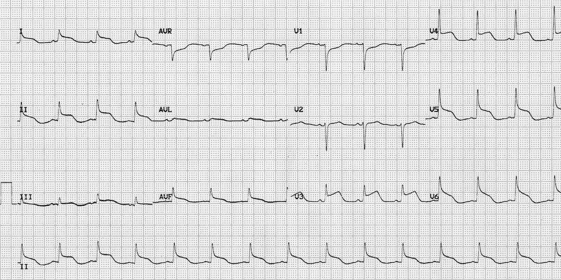
18-year old female with severe traumatic brain injury, ICP 40mmHg. Fluctuating BPs. Describe and interpret her ECG
Describe and interpret this ECG
ECG ANSWER and INTERPRETATION
- The ECG shows diffuse ST elevation.
- The morphology is atypical, there is no clear anatomical predominance for a vascular territory and no obvious reciprocal changes (except the inverted leads V1 and aVR).
- This is an example of a pseudo-infarction pattern due to raised intracranial pressure.
- Compare this with Quiz ECG 012 (raised ICP causing giant T wave inversions).
CLINICAL PEARLS
ECG changes with elevated ICP
Raised ICP is associated with certain characteristic ECG changes:
- Widespread giant T-wave inversions (“cerebral T waves”).
- QT prolongation.
- Bradycardia (the Cushing reflex – indicates imminent brainstem herniation).
Other possible ECG changes that may be seen:
- ST segment elevation / depression – this may mimic myocardial ischaemia or pericarditis.
- Increased U wave amplitude.
- Other rhythm disturbances: sinus tachycardia, junctional rhythms, premature ventricular contractions, atrial fibrillation.
In some cases, these ECG abnormalities may be associated with echocardiographic evidence of regional ventricular wall motion abnormality – so-called neurogenic stunned myocardium or neurogenic stress cardiomyopathy. The presumed mechanism is massive release of catecholamines, similar to Takutsubo syndrome.
This patient developed labile blood pressures and transient wall motion abnormalities plus these ECG changes during a sustained spike in her ICP. The ECG changes and wall motion abnormalities improved once her ICPs came under control.
References
Further Reading
- Wiesbauer F, Kühn P. ECG Mastery: Yellow Belt online course. Understand ECG basics. Medmastery
- Wiesbauer F, Kühn P. ECG Mastery: Blue Belt online course: Become an ECG expert. Medmastery
- Kühn P, Houghton A. ECG Mastery: Black Belt Workshop. Advanced ECG interpretation. Medmastery
- Rawshani A. Clinical ECG Interpretation ECG Waves
- Smith SW. Dr Smith’s ECG blog.
- Wiesbauer F. Little Black Book of ECG Secrets. Medmastery PDF
TOP 100 ECG Series
Emergency Physician in Prehospital and Retrieval Medicine in Sydney, Australia. He has a passion for ECG interpretation and medical education | ECG Library |

How do we know this is not pericarditis?